
Infectious Diseases — MCQs

On this page
Rose spot is associated with
True about diphtheria is -
Most common site of gastrointestinal TB:
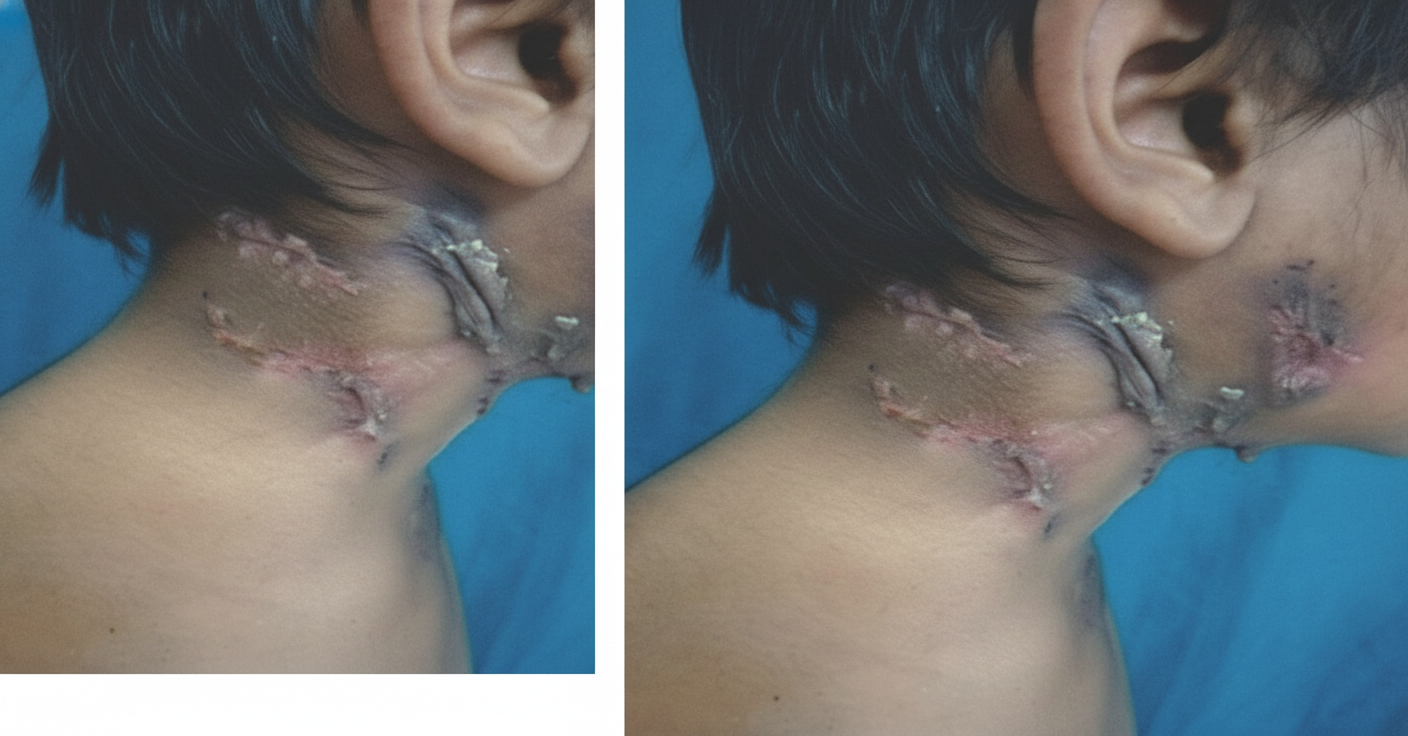
A lesion was seen on the face of a 42 year old patient as shown below. Which of the following would be ideal management for this condition?
HIV patient presented with diarrhea. On stool examination, acid-fast organisms (Isospora belli) were seen. What is the drug of choice in this patient?
After a renal transplant, what is the most common opportunistic infection?
Which of the following is not an AIDS defining illness?
Not an AIDS defining illness?
Mean transformation time for HIV to AIDS is:-
Match the following CSF findings with the most likely stage of syphilis: A. Normal CSF B. High protein, moderate pleocytosis C. High protein, high pleocytosis D. Normal protein, mild pleocytosis 1. Meningovascular syphilis 2. Primary syphilis 3. Tabes dorsalis 4. Meningeal syphilis
Practice by Chapter
Principles of Antimicrobial Therapy
Practice Questions
Fever of Unknown Origin
Practice Questions
HIV/AIDS and Related Infections
Practice Questions
Tuberculosis and Mycobacterial Diseases
Practice Questions
Tropical and Parasitic Infections
Practice Questions
Viral Infections (Hepatitis, Herpes, etc.)
Practice Questions
Healthcare-Associated Infections
Practice Questions
Fungal Infections
Practice Questions
Sepsis and Septic Shock
Practice Questions
Infection in Immunocompromised Hosts
Practice Questions
Emerging and Re-emerging Infections
Practice Questions
Antimicrobial Resistance
Practice Questions
Vaccination Principles
Practice Questions
Want unlimited practice?
Get full access to all questions, explanations, and performance tracking.
Scan to download app