
Infectious Diseases — MCQs

On this page
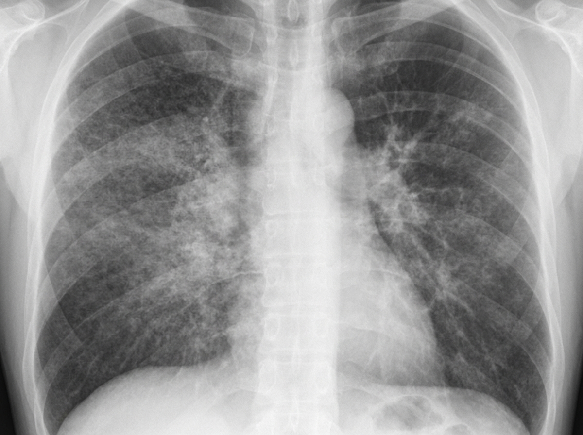
A young male presents with cough. The provided chest X-ray shows no known cardiac abnormality. Which of the following is most likely to be true about the patient?
What is the first sign of tetanus?
A 30-year-old male with a past history of otitis media and mastoiditis presented with severe throbbing headache, high-grade fever, nausea, vomiting, confusion, and gait disturbance. On examination, he was febrile, ataxic with nystagmus, and there was no neck rigidity. MRI brain with contrast revealed a brain abscess. Which of the following is the likely site of the brain abscess?
A patient with HIV is positive, with a viral load of 750,000 copies of HIV RNA/ml and a total CD4 count of 50. This patient is at an increased risk for several infectious diseases. For which of the following diseases does this patient have no added risk compared to an immunocompetent host?
What is a common cause of P.F?
A young man presents with a history of 6 kg weight loss in 3 months and on examination is found to have generalized lymph node enlargement. Which of the following is the LEAST likely diagnosis?
A 68-year-old woman develops new symptoms of burning when voiding. She has no fever, chills, or back discomfort. Her urinalysis reveals numerous white cells and bacteria. Which of the following medical comorbidities is most likely to coexist in this patient?
The tourniquet test is considered positive if the number of petechiae exceeds which threshold per square inch?
Which of the following statements about Pneumocystis pneumonia is true?
Which of the following is true about Lyme disease?
Practice by Chapter
Principles of Antimicrobial Therapy
Practice Questions
Fever of Unknown Origin
Practice Questions
HIV/AIDS and Related Infections
Practice Questions
Tuberculosis and Mycobacterial Diseases
Practice Questions
Tropical and Parasitic Infections
Practice Questions
Viral Infections (Hepatitis, Herpes, etc.)
Practice Questions
Healthcare-Associated Infections
Practice Questions
Fungal Infections
Practice Questions
Sepsis and Septic Shock
Practice Questions
Infection in Immunocompromised Hosts
Practice Questions
Emerging and Re-emerging Infections
Practice Questions
Antimicrobial Resistance
Practice Questions
Vaccination Principles
Practice Questions
Want unlimited practice?
Get full access to all questions, explanations, and performance tracking.
Scan to download app