
Infectious Diseases — MCQs

On this page
A 20-year-old male presents with fever, severe headache, vomiting, and photophobia. On examination, neck rigidity and Brudzinskis sign are positive. CT scan of the brain shows hydrocephalus. What is the most likely diagnosis?
A 58-year-old male presents with burning micturition. Prostatic examination is normal. Urinalysis shows >50 pus cells per high power field, but urine culture shows no growth. What is the most likely diagnosis?
A 56-year-old diabetic male presents with a complicated urinary tract infection and is found to be hypotensive. His blood pressure does not respond to intravenous fluids. Which of the following is the most appropriate antibiotic to manage his condition?
A patient underwent cardiac surgery and was admitted to the ICU. He later developed ventilator-associated pneumonia and was started on empirical antibiotics. A few hours later, the patient developed acute kidney injury. Serum procalcitonin was sent. Which of the following statements regarding procalcitonin is correct?
A 25-year-old sewage worker presents with fever for 1 week and weakness for 1 day. Laboratory evaluation reveals elevated bilirubin and decreased urine output. Conjunctival redness? What is the most likely diagnosis?
A patient with HIV develops tuberculosis. When should ART be initiated?
A patient with HIV is newly diagnosed with multidrug-resistant tuberculosis. Which of the following is the appropriate regimen, and what should the patient be monitored for?
According to WHO, which of the following is the recommended diagnostic test for spinal tuberculosis?
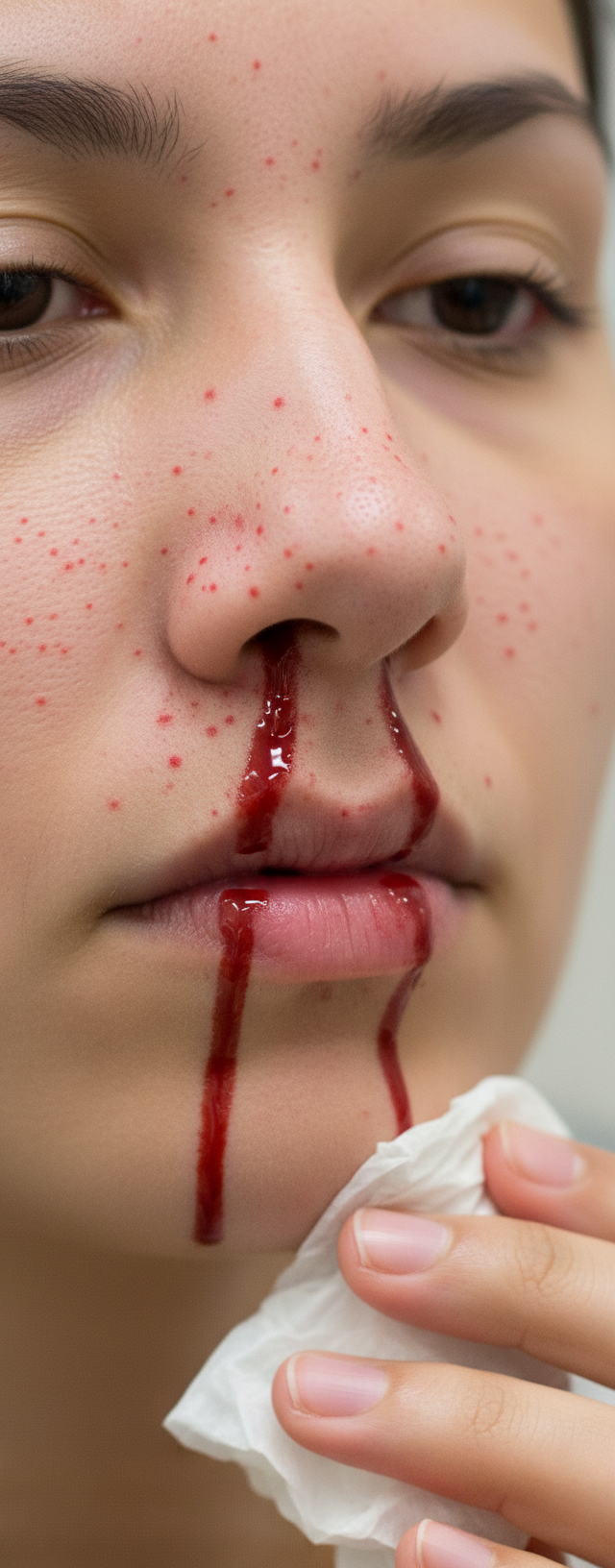
A 25 -year-male presents with high grade fever for 2 days. Vitals are pulse rate = 90 bpm and BP = 110/70 mm Hg. Labs were done and show positive NS-1 Antigen and platelet count = 40,000/cu.mm. The physical examination findings are shown below. What is the grade of severity of DHF?
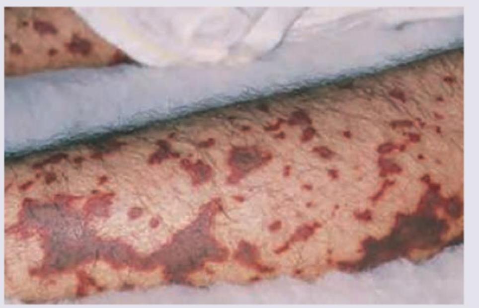
A 26-year-old male presented with fever and headache for 3 days. On 3rd day, his BP is $90 / 60 \mathrm{~mm} \mathrm{Hg}$ and examination revealed rashes on the legs as shown. What is the likely diagnosis?
Practice by Chapter
Principles of Antimicrobial Therapy
Practice Questions
Fever of Unknown Origin
Practice Questions
HIV/AIDS and Related Infections
Practice Questions
Tuberculosis and Mycobacterial Diseases
Practice Questions
Tropical and Parasitic Infections
Practice Questions
Viral Infections (Hepatitis, Herpes, etc.)
Practice Questions
Healthcare-Associated Infections
Practice Questions
Fungal Infections
Practice Questions
Sepsis and Septic Shock
Practice Questions
Infection in Immunocompromised Hosts
Practice Questions
Emerging and Re-emerging Infections
Practice Questions
Antimicrobial Resistance
Practice Questions
Vaccination Principles
Practice Questions
Want unlimited practice?
Get full access to all questions, explanations, and performance tracking.
Scan to download app