Ibalizumab was approved by the FDA in April 2018 for which condition?
Which statement is not true regarding dengue hemorrhagic fever?
One day after a casual sexual encounter with a bisexual man recently diagnosed as antibody-positive for human immunodeficiency virus (HIV), a patient is concerned about whether she may have become infected. A negative antibody titer is obtained. To test for seroconversion, when is the earliest repeat antibody testing should be scheduled after the sexual encounter?
What is the prophylactic antifungal drug commonly used in HIV patients?
What is the topical antifungal used in the treatment of Tinea corporis?
A man from Mexico presents with a clinical finding that, upon biopsy, reveals the presence of gram-negative bacilli. What is the most likely causative agent?
What is the cornerstone treatment for dengue fever with warning signs, in the absence of shock or hemorrhage?
Which of the following is a chronic complication of malaria?
Serology of a young man shows HBsAg, but HBeAg is negative with normal levels of AST and ALT. He is asymptomatic. What is the next line of management?
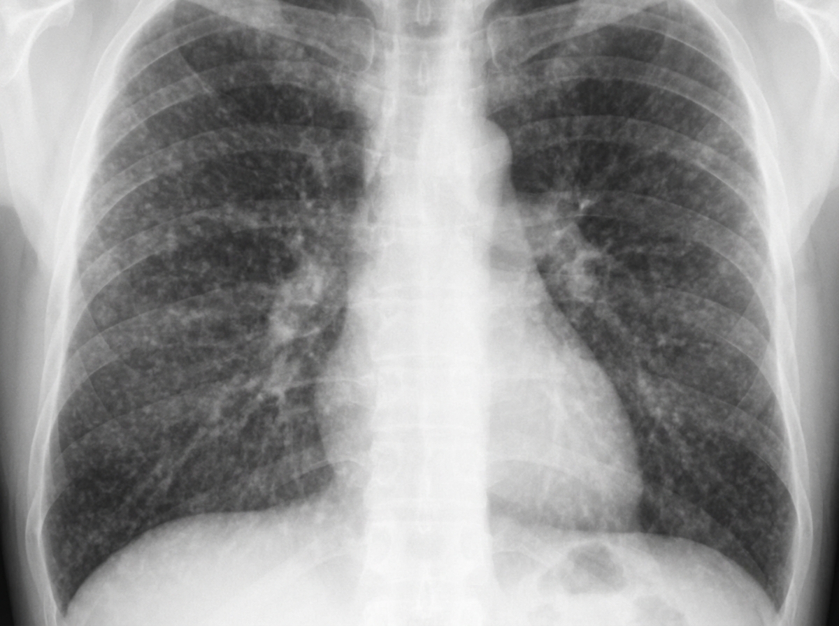
A 32-year-old woman seeking to become pregnant visits her physician for a pre-pregnancy examination. Routine prenatal laboratory testing demonstrates the following profile: HBsAg (-), anti-HBsAg (+), anti-HBcAg (-), anti-HBeAg (-), and HBV DNA (-). Which of the following likely represents the status of the patient?
Explanation: **Explanation:** **Ibalizumab** is a breakthrough medication in the management of **Human Immunodeficiency Virus (HIV)**. It is a humanized monoclonal antibody that acts as a **post-attachment inhibitor**. Unlike other entry inhibitors (like Maraviroc), Ibalizumab binds to the extracellular domain 2 of the **CD4+ T-cell receptor** [1]. This binding creates a conformational change that prevents the HIV-1 gp120 molecule from interacting with co-receptors (CCR5 or CXCR4), thereby blocking viral entry into the host cell. It was FDA-approved in 2018 specifically for **multidrug-resistant (MDR) HIV-1** in treatment-experienced adults failing their current antiretroviral regimen [1]. **Analysis of Incorrect Options:** * **Tuberculosis (TB):** TB is caused by *Mycobacterium tuberculosis* and is treated with antibiotics (RIPE regimen). Monoclonal antibodies are not currently a standard of care for TB. * **Leprosy:** Caused by *Mycobacterium leprae*, it is managed with Multi-Drug Therapy (MDT) involving Rifampicin, Dapsone, and Clofazimine. * **Malaria:** Caused by *Plasmodium* parasites, treatment involves antimalarials like Artemisinin-based combination therapies (ACTs) or Chloroquine. **High-Yield Clinical Pearls for NEET-PG:** * **Route of Administration:** Ibalizumab is administered **intravenous (IV)** every 14 days, making it unique among HIV medications. * **Mechanism:** It does not cause immunosuppression because it does not interfere with MHC II molecule binding or CD4-mediated immune responses. * **Indication:** Reserved for "Salvage Therapy" in patients with limited options due to extensive drug resistance [1]. * **Other Entry Inhibitors:** Remember **Enfuvirtide** (Fusion inhibitor - binds gp41) and **Maraviroc** (CCR5 antagonist) [1].
Explanation: **Explanation:** The diagnosis of **Dengue Hemorrhagic Fever (DHF)** is defined by a specific triad of clinical and laboratory findings. According to the WHO criteria, DHF is characterized by fever, bleeding manifestations, **thrombocytopenia**, and evidence of **plasma leakage** [1]. **Why Option A is the Correct Answer:** While shock is a severe complication of dengue, it is the defining feature of **Dengue Shock Syndrome (DSS)**, not DHF. DHF is categorized into four grades (I to IV). Grades I and II represent DHF without shock, while Grades III and IV represent DSS [1]. Therefore, shock is not a mandatory requirement for a diagnosis of DHF. **Analysis of Incorrect Options:** * **B. Thrombocytopenia:** This is a mandatory criterion for DHF. A platelet count of **<100,000 cells/mm³** is required for the diagnosis. * **C. Hepatomegaly:** Enlargement of the liver is a common clinical finding in DHF and is included in the WHO diagnostic criteria as a supportive clinical sign [1]. * **D. Plasma Leakage:** This is the **hallmark** that differentiates DHF from classic Dengue Fever. It is evidenced by a >20% rise in hematocrit, pleural effusion, or ascites. **High-Yield Clinical Pearls for NEET-PG:** * **Critical Phase:** Usually occurs during days 3–7 of illness (defervescence), which is when plasma leakage and shock are most likely to develop [1]. * **Tourniquet Test:** A positive test (≥10 petechiae per square inch) suggests capillary fragility, common in DHF [1]. * **Most common cause of death:** Hypovolemic shock due to plasma leakage. * **Vector:** *Aedes aegypti* (Day biter).
Explanation: ### Explanation **Correct Option: C (6 to 12 weeks)** The primary concept here is the **"Window Period."** This is the interval between the initial HIV infection and the time when the body produces enough antibodies to be detected by standard screening tests (ELISA) [1]. While modern 4th-generation assays (which detect both p24 antigen and antibodies) can detect infection as early as 2–3 weeks, the traditional **antibody-only seroconversion** typically occurs within **6 to 12 weeks** in the vast majority of patients. By 3 months (12 weeks), more than 95% of individuals will have detectable antibodies [3]. Therefore, to reliably confirm or rule out seroconversion via antibody testing, the earliest recommended window starts at 6 weeks [3]. **Analysis of Incorrect Options:** * **A (1 to 2 weeks):** This is too early for an antibody response. Only HIV-RNA (viral load) via PCR might be detectable at this stage [2]. * **B (3 to 4 weeks):** While a 4th-generation p24 antigen test might be positive, an **antibody-only** titer (as specified in the question) is often still negative during this period. * **D (12 to 15 weeks):** While testing at this stage is highly accurate, it is not the *earliest* time to schedule a repeat test. 6 to 12 weeks is the standard diagnostic window [3]. **NEET-PG High-Yield Pearls:** 1. **Order of Detection:** Viral RNA (10–12 days) → p24 Antigen (14–16 days) → Antibodies (3–12 weeks). 2. **Screening Test:** ELISA (High sensitivity) [1]. 3. **Confirmatory Test:** Western Blot (detects antibodies to specific proteins like gp41, gp120, p24) [1]. *Note: Newer algorithms use HIV-1/2 differentiation assays.* 4. **Post-Exposure Prophylaxis (PEP):** Must be started within **72 hours** of exposure and continued for 28 days (Tenofovir + Emtricitabine + Dolutegravir/Raltegravir).
Explanation: In the context of HIV/AIDS, antifungal prophylaxis is primarily indicated for patients with severe immunosuppression (CD4 count <100 cells/µL) to prevent endemic mycoses. [1] **Why Itraconazole is Correct:** **Itraconazole** is the drug of choice for the primary prophylaxis of **Histoplasmosis** and **Penicilliosis** (Talaromycosis) in HIV-infected individuals living in endemic geographical areas. It is preferred due to its broad spectrum against these specific dimorphic fungi and its established safety profile in long-term HIV management. While Fluconazole is more common for Cryptococcal prophylaxis [2], Itraconazole remains the standard answer for general "prophylactic antifungal" questions in many medical exams focusing on endemic regions. **Analysis of Incorrect Options:** * **B. Posaconazole:** While highly effective, it is typically reserved for prophylaxis in patients with hematological malignancies and severe Neutropenia (e.g., GVHD), rather than routine HIV care, due to its high cost. * **C. Terconazole:** This is a topical antifungal used exclusively for the treatment of vulvovaginal candidiasis; it has no role in systemic prophylaxis. * **D. Voriconazole:** This is the gold standard for the *treatment* of Invasive Aspergillosis. It is rarely used for prophylaxis in HIV because of its significant drug-drug interactions with Protease Inhibitors and NNRTIs via the CYP450 enzyme system. **High-Yield Clinical Pearls for NEET-PG:** * **Histoplasmosis Prophylaxis:** Indicated if CD4 <150 cells/µL in endemic areas (Itraconazole 200mg/day). [5] * **Cryptococcal Prophylaxis:** Primary prophylaxis is generally not recommended unless ART is unavailable; however, if started, **Fluconazole** is used. [4] * **Pneumocystis jirovecii (PJP):** The most common opportunistic infection; prophylaxis is with **TMP-SMX** (Cotrimoxazole) when CD4 <200 cells/µL. [3] * **Drug of Choice for Induction:** Amphotericin B is used for induction therapy in most systemic fungal infections in HIV, followed by Itraconazole or Fluconazole for maintenance.
Explanation: **Explanation:** **Tinea corporis** (ringworm) is a superficial dermatophyte infection of the glabrous skin [1]. The primary goal of treatment for localized, non-extensive lesions is the use of **topical antifungal agents** [1][2]. **1. Why Tolnaftate is Correct:** **Tolnaftate** is a topical thiocarbamate antifungal. It works by inhibiting the enzyme **squalene epoxidase**, which is essential for ergosterol synthesis in fungal cell membranes. It is highly effective against dermatophytes (Trichophyton, Microsporum, and Epidermophyton) and is a standard over-the-counter topical treatment for uncomplicated Tinea corporis. **2. Why the Other Options are Incorrect:** * **Itraconazole (B) and Fluconazole (C):** These are systemic triazoles [2]. While highly effective, they are reserved for extensive disease, tinea capitis, tinea unguium (onychomycosis), or cases refractory to topical therapy [2]. In the context of "topical antifungal used," these do not fit as they are primarily administered orally [2]. * **Griseofulvin (D):** This is an oral fungistatic agent that binds to tubulin, interfering with microtubule function. It is never used topically and is primarily indicated for Tinea capitis in children. **Clinical Pearls for NEET-PG:** * **First-line Topical Agents:** Terbinafine (Allylamine) is often considered more effective than Tolnaftate or Azoles due to its fungicidal properties. * **Steroid Warning:** Avoid topical steroids (e.g., Clobetasol) in Tinea; it leads to **Tinea Incognito**, where the classic annular morphology is lost, but the infection worsens [1]. * **Diagnosis:** The gold standard for bedside diagnosis is **KOH mount**, showing translucent, septate branching hyphae. * **Drug of Choice (Systemic):** Oral Terbinafine is currently the preferred systemic agent for dermatophytosis over Griseofulvin.
Explanation: The clinical presentation and biopsy findings point toward **Rhinoscleroma**, a chronic granulomatous condition of the upper respiratory tract. **1. Why Klebsiella rhinoscleromatis is correct:** *Klebsiella pneumoniae* subspecies *rhinoscleromatis* is a **gram-negative bacillus** that causes Rhinoscleroma. It is endemic in Central America (including Mexico), Africa, and the Middle East. The hallmark histological finding is the **Mikulicz cell**—a large, foamy macrophage containing the causative gram-negative bacilli. The disease typically progresses through three stages: catarrhal, proliferative (granulomatous), and cicatricial (scarring). **2. Why the other options are incorrect:** * **Mycobacterium leprae (B):** While it causes chronic granulomatous lesions (Leprosy), it is an **acid-fast bacillus**, not gram-negative [1]. Histology shows Virchow cells (lepra cells). Treatment involves multidrug therapy containing rifampicin [1]. * **Acinetobacter baumannii (C):** This is a gram-negative coccobacillus, but it typically causes acute opportunistic infections like ventilator-associated pneumonia or wound infections in ICU settings, not chronic granulomatous nasal lesions. * **Treponema pallidum (D):** The causative agent of Syphilis is a **spirochete**. While it can cause nasal gummas (Tertiary Syphilis), it is not classified as a gram-negative bacillus and is best visualized via dark-field microscopy or silver stains (Warthin-Starry). Other treponemal infections like yaws also present with destructive granulomatous changes that heal with scarring [2]. **High-Yield NEET-PG Pearls:** * **Mikulicz Cells:** Pathognomonic for Rhinoscleroma (foamy macrophages with bacilli). * **Russell Bodies:** Eosinophilic inclusions (representing immunoglobulins) often seen in the plasma cells of these lesions. * **Treatment:** Long-term antibiotics (Streptomycin and Tetracycline/Ciprofloxacin) are usually required. * **Warthin-Starry Stain:** Can be used to highlight the bacilli within the Mikulicz cells.
Explanation: The cornerstone of management for dengue fever with warning signs is **Intravenous (IV) fluid therapy**. The pathophysiology of severe dengue is characterized by increased capillary permeability leading to plasma leakage. This results in hemoconcentration and potential hypovolemic shock [1]. Administering isotonic crystalloids (like Normal Saline or Ringer’s Lactate) counteracts this leakage, maintains intravascular volume, and prevents progression to Dengue Shock Syndrome (DSS) [1]. **Analysis of Incorrect Options:** * **A. Platelet transfusion:** This is not indicated based on platelet count alone. It is reserved for patients with significant bleeding or very low counts (<10,000/mm³) with high risk of hemorrhage. Prophylactic transfusion does not improve outcomes. * **C. Cryoglobulins:** These have no role in the management of dengue. Cryoprecipitate may be used in rare cases of disseminated intravascular coagulation (DIC), but it is not a primary treatment. * **D. Antivirals:** There are currently no approved or effective antiviral drugs for the treatment of Dengue virus; management remains purely supportive. **High-Yield Clinical Pearls for NEET-PG:** * **Warning Signs:** Abdominal pain/tenderness, persistent vomiting, mucosal bleed, lethargy, liver enlargement >2cm, and a rapid increase in Hct with a rapid decrease in platelet count. * **Fluid Choice:** Isotonic crystalloids are the first choice. Colloids are reserved for refractory shock. * **Critical Period:** Usually occurs between days 3–7 of illness during the "defervescence" phase (when fever drops) [1]. This is when plasma leakage is most severe. * **Avoid:** NSAIDs (like Aspirin/Ibuprofen) due to the risk of bleeding; use Paracetamol for fever.
Explanation: The correct answer is Both nephrotic syndrome and splenomegaly because chronic or repeated exposure to malaria parasites leads to persistent immune activation and structural organ changes. 1. Nephrotic Syndrome: This is a classic chronic complication specifically associated with Plasmodium malariae. It results from the deposition of immune complexes in the glomerular basement membrane (Quartan Malarial Nephropathy). Unlike the acute kidney injury seen in P. falciparum, this is a chronic, progressive condition that often responds poorly to antimalarial therapy. 2. Splenomegaly: Chronic or recurrent malaria leads to persistent congestion and hyperplasia of the reticuloendothelial system [1]. A specific chronic manifestation is Hyperreactive Malarial Splenomegaly (HMS), formerly known as Tropical Splenomegaly Syndrome. It is characterized by massive splenomegaly, high titers of anti-malarial antibodies, and hypergammaglobulinemia (IgM). Why other options are incorrect: * Pneumonia: While pulmonary edema and ARDS are severe acute complications of P. falciparum malaria, pneumonia is typically a secondary bacterial superinfection rather than a direct chronic complication of the malaria parasite itself. High-Yield Clinical Pearls for NEET-PG: * HMS Treatment: Long-term antimalarial chemoprophylaxis (e.g., Proguanil or Chloroquine) is the treatment of choice for Hyperreactive Malarial Splenomegaly. * Blackwater Fever: An acute complication involving massive intravascular hemolysis and hemoglobinuria, usually associated with P. falciparum and quinine use. * Recrudescence vs. Relapse: P. falciparum causes recrudescence (sub-optimal treatment) [1], while P. vivax and P. ovale cause relapse due to hypnozoites in the liver [1].
Explanation: ### Explanation The patient in this scenario is an **Inactive HBsAg Carrier**. This clinical state is characterized by the presence of HBsAg for >6 months, the absence of HBeAg (HBeAg negative), the presence of Anti-HBe, low HBV DNA levels (<2,000 IU/mL), and persistently normal ALT/AST levels [1]. **1. Why "Wait and Watch" is correct:** In an asymptomatic patient with normal liver enzymes and no evidence of active viral replication (HBeAg negative), the risk of liver damage is currently very low. According to AASLD and EASL guidelines, antiviral therapy is only indicated if there is evidence of active chronic hepatitis (elevated ALT and high HBV DNA) or cirrhosis. For inactive carriers, the standard management is **long-term monitoring** (serial ALT and HBsAg testing) to detect potential reactivation [1]. **2. Why the other options are incorrect:** * **Antiviral Therapy:** Not indicated because the ALT is normal and the patient is asymptomatic. Unnecessary treatment increases the risk of drug resistance and side effects. * **Immunoglobulins (HBIG):** These are used for post-exposure prophylaxis (e.g., needle-stick injury) or to prevent reinfection post-liver transplant. They have no role in treating established chronic HBsAg carriers. * **Liver Transplant:** This is the treatment of choice for end-stage liver disease (decompensated cirrhosis) or fulminant hepatic failure, neither of which is present here. ### NEET-PG High-Yield Pearls * **HBeAg** is a marker of **active viral replication** and high infectivity [1]. * The first marker to appear after infection is **HBsAg**. * The first antibody to appear is **Anti-HBc (IgM)**; it is the only marker present during the **"Window Period."** * **Treatment Criteria:** Generally initiated if ALT >2x Upper Limit of Normal AND HBV DNA >2,000 IU/mL (in HBeAg negative) or >20,000 IU/mL (in HBeAg positive). * **Drug of Choice:** Tenofovir or Entecavir are preferred first-line agents due to high potency and low resistance.
Explanation: ### Explanation The patient’s serological profile indicates immunity acquired through **vaccination**. **1. Why the Correct Answer is Right:** The presence of **anti-HBsAg (+)** in the absence of other markers is the hallmark of successful immunization. * **HBsAg (-):** Indicates no active infection [1]. * **Anti-HBsAg (+):** Indicates protective immunity. * **Anti-HBcAg (-):** This is the "crucial differentiator." Anti-HBc (Antibody to Core antigen) is only produced in response to an actual viral infection (natural infection). Since vaccines contain only the recombinant surface protein (HBsAg) and no core components, a vaccinated person will be **Anti-HBc negative** [1]. **2. Why the Other Options are Wrong:** * **Option A (Carrier):** A chronic carrier would be **HBsAg (+)** for more than 6 months and **Anti-HBc (+)** (IgG) [1]. * **Option C (Window Period):** During the "window period," HBsAg has disappeared but Anti-HBs has not yet appeared. The only marker typically positive is **Anti-HBc IgM** [1]. * **Option D (Highly Transmissible):** High infectivity is characterized by the presence of **HBsAg (+)** and **HBeAg (+)**, along with high levels of **HBV DNA**. **3. NEET-PG High-Yield Pearls:** * **Natural Infection (Recovered):** HBsAg (-), **Anti-HBc (+)**, Anti-HBs (+) [1]. * **Vaccinated:** HBsAg (-), **Anti-HBc (-)**, Anti-HBs (+). * **HBeAg:** Represents active viral replication and high infectivity. * **Anti-HBs Titer:** A level of **≥10 mIU/mL** after a 3-dose vaccine series is considered protective. * **Isolated Anti-HBc (+):** Can represent a "resolved" infection where Anti-HBs has waned, a "window period," or a false positive.
Principles of Antimicrobial Therapy
Practice Questions
Fever of Unknown Origin
Practice Questions
HIV/AIDS and Related Infections
Practice Questions
Tuberculosis and Mycobacterial Diseases
Practice Questions
Tropical and Parasitic Infections
Practice Questions
Viral Infections (Hepatitis, Herpes, etc.)
Practice Questions
Healthcare-Associated Infections
Practice Questions
Fungal Infections
Practice Questions
Sepsis and Septic Shock
Practice Questions
Infection in Immunocompromised Hosts
Practice Questions
Emerging and Re-emerging Infections
Practice Questions
Antimicrobial Resistance
Practice Questions
Vaccination Principles
Practice Questions
Get full access to all questions, explanations, and performance tracking.
Start For Free