
Infectious Diseases — MCQs

On this page
A 20-year-old male goes swimming and after a few days develops diffuse itching with rashes over his body. Several weeks later, he develops lancinating pain down his legs and in all his toes. Within a few days, he develops paraparesis and problems with bowel and bladder control, resulting in urinary retention. What is your initial diagnostic approach for this patient?
What drug is used for the chemoprophylaxis of leptospirosis?
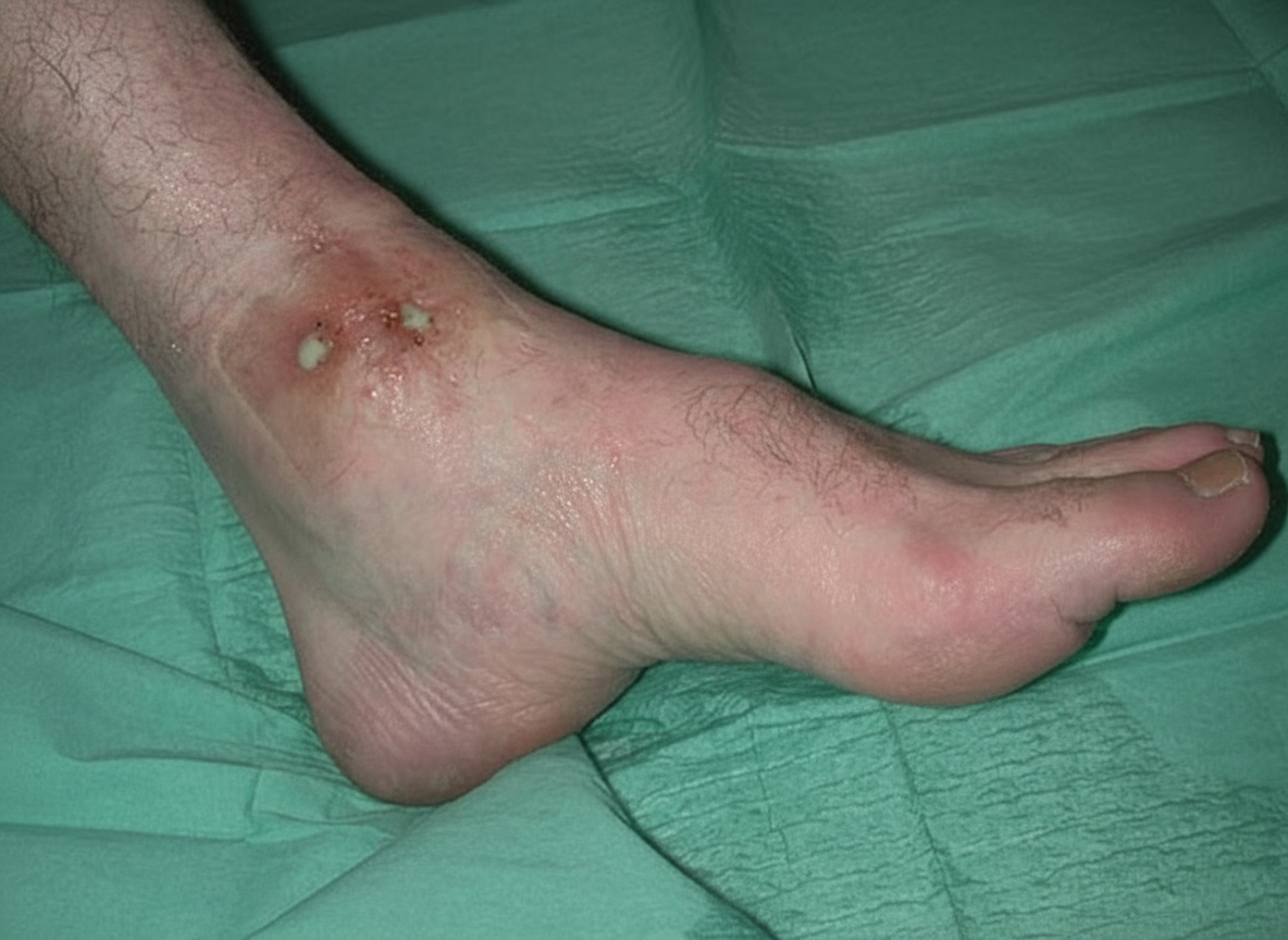
What is the most likely diagnosis in a patient from Mauritania whose foot developed these abnormalities over several years?
Which of the following is an initial presentation of HIV infection?
A lung biopsy of an HIV-positive patient shows intra-nuclear basophilic inclusion bodies with a perinuclear halo. The patient's CD4 count was less than 100 at the time of diagnosis. What is the probable cause?
A healthcare worker sustains a needle-prick injury from a patient with HIV. What is the immediate management or treatment required?
A 65-year-old man presents with fever, severe headache, and nuchal rigidity. Physical examination shows a Glasgow coma score of 7. Lumbar puncture reveals cloudy cerebrospinal fluid (CSF) with 1200 neutrophils/mm3, elevated protein, and decreased glucose. Which of the following is the most probable etiologic agent of this condition?
What is the most common late central nervous system complication of HIV infection?
A 23-year-old patient, studying abroad, visited his hometown and developed a high-grade fever (103°F) after 3 days. He took acetaminophen and subsequently complained of myalgias, cough, and mild shortness of breath. A nasopharyngeal swab tested positive for COVID-19. Which of the following statements regarding the patient's next steps in management is false?
What is the next step in the management of a patient with a positive sputum smear for tuberculosis but a negative chest X-ray?
Practice by Chapter
Principles of Antimicrobial Therapy
Practice Questions
Fever of Unknown Origin
Practice Questions
HIV/AIDS and Related Infections
Practice Questions
Tuberculosis and Mycobacterial Diseases
Practice Questions
Tropical and Parasitic Infections
Practice Questions
Viral Infections (Hepatitis, Herpes, etc.)
Practice Questions
Healthcare-Associated Infections
Practice Questions
Fungal Infections
Practice Questions
Sepsis and Septic Shock
Practice Questions
Infection in Immunocompromised Hosts
Practice Questions
Emerging and Re-emerging Infections
Practice Questions
Antimicrobial Resistance
Practice Questions
Vaccination Principles
Practice Questions
Want unlimited practice?
Get full access to all questions, explanations, and performance tracking.
Scan to download app