
Infectious Diseases — MCQs

On this page
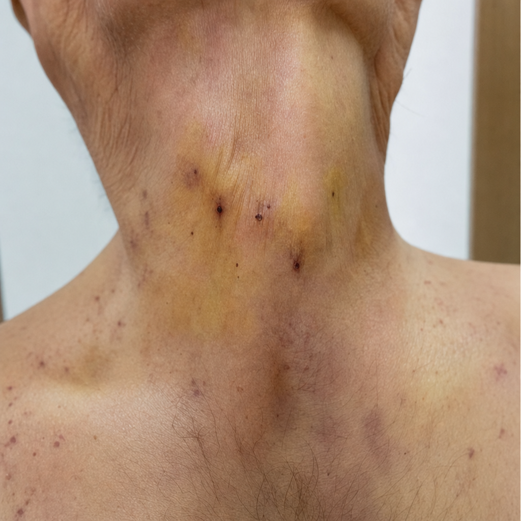
Which one of the following is typically associated with the given clinical finding?
A 24-year-old patient complains of diarrhea, vomiting, and abdominal pain aggravated on ingestion of food for 1 week. Stool microscopy reveals findings suggestive of parasitic infection. What is the appropriate treatment for this condition?

Hemorrhagic rashes are typically seen in which of the following conditions?
A patient presents with arthralgia, a rash, lymphadenopathy, and pneumonia, but no fever. Which of the following conditions is most likely given these symptoms?
A 31-year-old Haitian woman is evaluated for infertility. Pelvic examination shows a markedly enlarged vulva, inguinal lymph node enlargement, and rectal stricture. Biopsy of an inguinal lymph node reveals necrotizing granulomas, neutrophilic infiltrates, and inclusion bodies within macrophages. Which of the following is the most likely etiology of infertility in this patient?
A 45-year-old male patient presents with cough and diarrhea for the past 3 weeks. He is diagnosed as HIV positive with Tuberculosis. What should be the next line of management?
In fever of unknown origin, how many blood samples should ideally be drawn?
Trophic ulcers are seen in which of the following conditions?
What is occult hepatitis B?
Which of the following is the most common oral manifestation of infectious mononucleosis?
Practice by Chapter
Principles of Antimicrobial Therapy
Practice Questions
Fever of Unknown Origin
Practice Questions
HIV/AIDS and Related Infections
Practice Questions
Tuberculosis and Mycobacterial Diseases
Practice Questions
Tropical and Parasitic Infections
Practice Questions
Viral Infections (Hepatitis, Herpes, etc.)
Practice Questions
Healthcare-Associated Infections
Practice Questions
Fungal Infections
Practice Questions
Sepsis and Septic Shock
Practice Questions
Infection in Immunocompromised Hosts
Practice Questions
Emerging and Re-emerging Infections
Practice Questions
Antimicrobial Resistance
Practice Questions
Vaccination Principles
Practice Questions
Want unlimited practice?
Get full access to all questions, explanations, and performance tracking.
Scan to download app