
Infectious Diseases — MCQs

On this page
A 40-year-old man returns from rural Cambodia with fever and severe thrombocytopenia (platelet count 15,000/μL). Peripheral smear shows ring forms with multiple infections per RBC and appliqué forms. Rapid diagnostic test is positive for Plasmodium falciparum HRP2. What is the most appropriate first-line treatment?
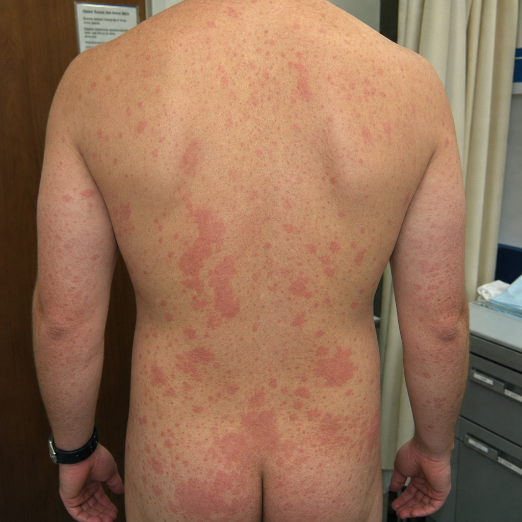
A 42-year-old man presents with fever, severe joint pains in small joints of hands and feet, and maculopapular rash for 5 days. He returned from Caribbean islands 10 days ago. RT-PCR confirms alphavirus infection. What is the most appropriate management?
A rash appeared 9 hours after a scuba dive. What is the diagnosis?
Which of the following statements regarding Pneumocystis jirovecii pneumonia (PCP) is/are true? 1. Bronchoscopy with bronchoalveolar lavage (BAL) is the mainstay of diagnosis for PCP. 2. Pneumatoceles are seen in all cases of PCP. 3. A CD4 count < 350 cells/µL is an indication for prophylaxis against PCP. 4. Trimethoprim-sulfamethoxazole is the drug of choice for prophylaxis.
The tourniquet test is used for monitoring patients with which of the following conditions?
What is the recommended dose of tetanus toxoid to prevent maternal and neonatal tetanus for a pregnant woman whose previous immunization status is not known?
In a person with HIV-1 infection, which of the following is the most predictive of the patient's prognosis?
In Human Immunodeficiency Virus (HIV) infection, diffuse lymphadenopathy in a person who is clinically well is usually a sign of which of the following?
The most severe form of respiratory complication in influenza virus infection is:
What is the percentage of coincidence between a sore throat and acute rheumatic fever?
Practice by Chapter
Principles of Antimicrobial Therapy
Practice Questions
Fever of Unknown Origin
Practice Questions
HIV/AIDS and Related Infections
Practice Questions
Tuberculosis and Mycobacterial Diseases
Practice Questions
Tropical and Parasitic Infections
Practice Questions
Viral Infections (Hepatitis, Herpes, etc.)
Practice Questions
Healthcare-Associated Infections
Practice Questions
Fungal Infections
Practice Questions
Sepsis and Septic Shock
Practice Questions
Infection in Immunocompromised Hosts
Practice Questions
Emerging and Re-emerging Infections
Practice Questions
Antimicrobial Resistance
Practice Questions
Vaccination Principles
Practice Questions
Want unlimited practice?
Get full access to all questions, explanations, and performance tracking.
Scan to download app