
Infectious Diseases — MCQs

On this page
In chronic Hepatitis B (HBV) infection, the presence of HBeAg suggests which of the following?
What is the most common pulmonary manifestation in AIDS?
Which of the following laboratory markers characterizes the recumbent stage of Hepatitis B?
What is the true statement regarding the treatment of hepatic amoebiasis?
A 55-year-old diabetic female patient presented with facial pain, numbness, conjunctival suffusion, and blurring of vision for 6 days. She was managed conservatively and discharged. After a week, she presented again with bilateral proptosis, chemosis, vision loss, and ophthalmoplegia, along with a characteristic lesion on the nose. A smear was prepared from the nasal lesion. What is the most appropriate initial management for a suspected fungal infection causing these symptoms?
Fever, clubbing, and Osler's nodes are characteristic findings in which of the following conditions?
A 40-year-old man presents with fever, weight loss, and cough. His Mantoux test result is 18 x 19 mm, and sputum cytology is negative for Acid-Fast Bacilli (AFB). What is the most likely condition?
A 68-year-old woman presents with new symptoms of burning when voiding. She has no fever, chills, or back discomfort. Her urinalysis reveals numerous white cells and bacteria. Which of the following medical comorbidities is most likely to coexist in this patient?
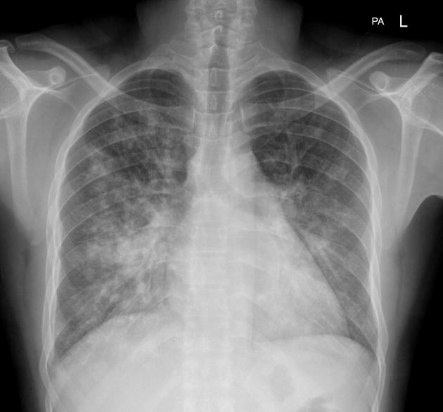
A 35-year-old male patient with a known case of HIV on Antiretroviral Therapy complains of fever, dyspnea, dry cough, and weight loss for the past 3 months. The chest X-ray of the patient is as below. What is your diagnosis?
A 22-year-old male military recruit complains of a headache and stiff neck. He is examined, blood is drawn, and a lumbar puncture is performed. The glucose in the CSF is 100 mg/dL and the serum glucose is 120 mg/dL. The CSF shows 3 lymphocytes and 0 neutrophils/microliter. Which of the following conclusions concerning the interpretation of these findings is most accurate?
Practice by Chapter
Principles of Antimicrobial Therapy
Practice Questions
Fever of Unknown Origin
Practice Questions
HIV/AIDS and Related Infections
Practice Questions
Tuberculosis and Mycobacterial Diseases
Practice Questions
Tropical and Parasitic Infections
Practice Questions
Viral Infections (Hepatitis, Herpes, etc.)
Practice Questions
Healthcare-Associated Infections
Practice Questions
Fungal Infections
Practice Questions
Sepsis and Septic Shock
Practice Questions
Infection in Immunocompromised Hosts
Practice Questions
Emerging and Re-emerging Infections
Practice Questions
Antimicrobial Resistance
Practice Questions
Vaccination Principles
Practice Questions
Want unlimited practice?
Get full access to all questions, explanations, and performance tracking.
Scan to download app