
Infectious Diseases — MCQs

On this page
A diabetic patient presents with fungal infection of the sinuses and peri-orbital region with significant visual impairment. What is the best drug for treatment in this patient?
All the following are characteristics of AIDS when CD4 cell count drops below 100 cells, EXCEPT:
Reactivation tuberculosis is usually seen where?
A 40-year-old paddy farmer presented with fever, chills, headache, and myalgias for 2 days. The patient also complained of acute onset of cough, shortness of breath, and a few episodes of hemoptysis. On examination, scleral icterus was present. There is a history of a minor lower limb injury while working in the fields, which were infested with rats. Lab findings revealed anemia, leukocytosis, deranged RFTs, prolonged PT and aPTT, increased serum bilirubin and alkaline phosphatase levels. Blood cultures, dark field microscopy, cultures, and MAT test were performed. Which electrolyte derangement will be most likely seen in the above condition?
What is the most common ophthalmic lesion in AIDS?
Which viral infections are associated with hemolysis?
Most common central nervous system (CNS) manifestation of HIV infection is:
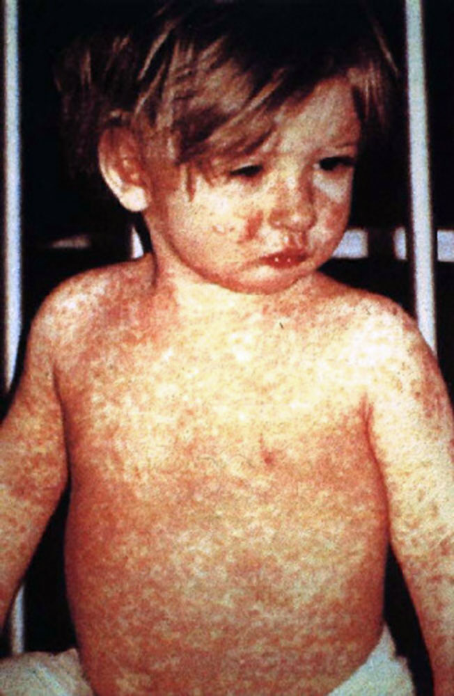
A 60-year-old unvaccinated patient presents to the OPD with fever and red eyes. On examination, a specific type of rash is observed on the body. What is the most likely complication expected with this condition?
A patient with AIDS presents with an acute episode of diarrhea. Stool examination reveals an oval structure, 8 to 9 micrometers in diameter, which is acid-fast and fluoresces blue under ultraviolet light. What is the drug of choice for this patient?
What is true about amoebic liver abscess?
Practice by Chapter
Principles of Antimicrobial Therapy
Practice Questions
Fever of Unknown Origin
Practice Questions
HIV/AIDS and Related Infections
Practice Questions
Tuberculosis and Mycobacterial Diseases
Practice Questions
Tropical and Parasitic Infections
Practice Questions
Viral Infections (Hepatitis, Herpes, etc.)
Practice Questions
Healthcare-Associated Infections
Practice Questions
Fungal Infections
Practice Questions
Sepsis and Septic Shock
Practice Questions
Infection in Immunocompromised Hosts
Practice Questions
Emerging and Re-emerging Infections
Practice Questions
Antimicrobial Resistance
Practice Questions
Vaccination Principles
Practice Questions
Want unlimited practice?
Get full access to all questions, explanations, and performance tracking.
Scan to download app