
Critical Care — MCQs

On this page
Refractory Septic shock is defined as?
All of the following can cause Pulseless Electrical Activity (PEA) except
What is the first-line fluid to be administered in a patient presenting with acute hemorrhagic shock?
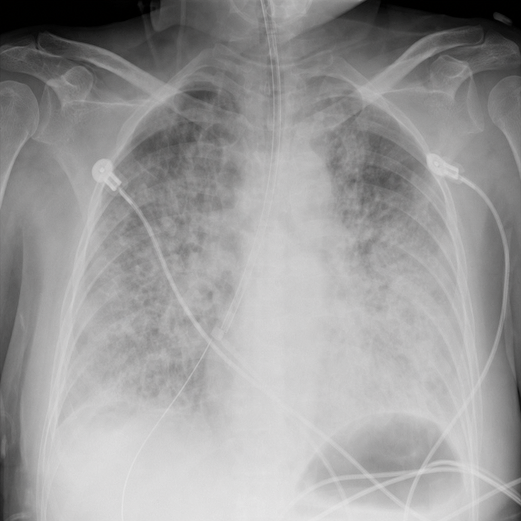
A 64-year-old woman is admitted to the ICU with the clinical diagnosis of acute respiratory distress syndrome (ARDS) secondary to pneumonia. She requires intubation and mechanical ventilation. On the second ICU day, she is difficult to ventilate, requiring increased airway pressures. On physical examination, vital signs are: pulse 159 bpm; temperature 100degF; blood pressure 90/56 mm Hg. Lung exam reveals diffuse crackles, and the patient has a palpable crunch on exam of her chest wall and abdomen. Chest radiograph is shown below.. What will you do next?
What is the maximum possible score in the APACHE II scoring system?
Which of the following is the best method to assess the adequacy of fluid replacement?
Which is the best initial fluid for resuscitation during the shock state:
To prevent ventilator associated pneumonia, the most effective and evidence based results are seen with which of the following for critically ill patients:
Which of the following is a feature of crush syndrome -
A patient presents with multiple fractures. He develops respiratory distress and dies after few days. CT brain shows petechial hemorrhage. Most likely diagnosis is:
Practice by Chapter
Shock Syndromes and Management
Practice Questions
Acute Respiratory Distress Syndrome
Practice Questions
Mechanical Ventilation Principles
Practice Questions
Hemodynamic Monitoring
Practice Questions
Nutrition in Critical Illness
Practice Questions
Sedation and Analgesia in ICU
Practice Questions
Multi-organ Dysfunction Syndrome
Practice Questions
Acid-Base and Electrolyte Disturbances
Practice Questions
Toxicologic Emergencies
Practice Questions
Neurological Emergencies in ICU
Practice Questions
Renal Replacement Therapy
Practice Questions
End-of-Life Care in ICU
Practice Questions
Want unlimited practice?
Get full access to all questions, explanations, and performance tracking.
Scan to download app