
Critical Care — MCQs

On this page
In a septic shock patient who remains hypotensive despite adequate fluid resuscitation, what would be the next drug of choice?
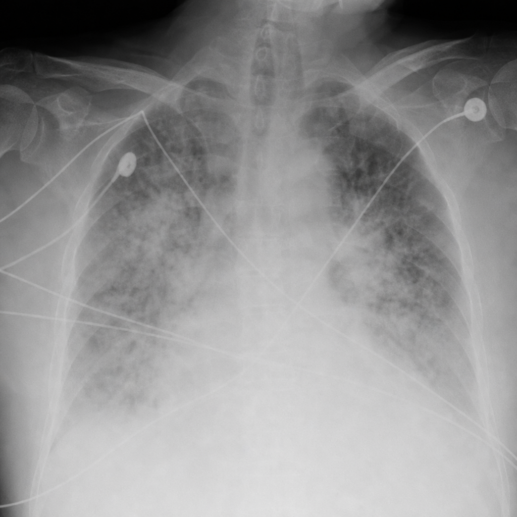
A 65-year-old alcoholic is admitted to the ICU with diagnosis of acute pancreatitis. After 48 hours he is unconscious and has the following findings: $\mathrm{sPO}_{2}=$ $60 \%, \mathrm{pO}_{2}=60 \mathrm{~mm} \mathrm{Hg}, \mathrm{pCO}_{2}=30 \mathrm{~mm} \mathrm{Hg}$ and $\mathrm{HR}=$ 120 bpm . CXR was performed. Diagnosis is?
Which of the following are components of SOFA scoring system? I. PaO_2 / FiO_2 ratio II. Mean arterial pressure III. Glasgow coma scale IV. Prothrombin Time with INR Select the correct answer using the code given below :
Which of the following are correct in respect of Systemic Inflammatory Response Syndrome (SIRS)? 1. It is caused by the release of lipopolysaccharide endotoxin from dying E. coli bacteria. 2. It is same as bacteraemia. 3. It results in Multiple Organ Dysfunction Syndrome (MODS). 4. White cell counts of more than 12 × 10^9/litre are present. Select the answer using the code given below.
Consider the following statements : Systemic Inflammatory Response Syndrome is characterised by 1. Temperature either above 38°C or below 36°C. 2. Heart rate less than 80/minute. 3. Tachypnoea > 20/min. 4. Leucocyte count > 4 x 10⁹/L. Which of the statements given above are correct ?
Systemic Inflammatory Response Syndrome (SIRS) diagnostic criteria include the following except
Which of the following is NOT a feature of Systemic Inflammatory Response Syndrome?
A 25 year old lady underwent exploratory laparotomy for bowel injury which happened while she underwent medical termination of pregnancy 2 days back. 24 hours after exploratory laparotomy her pulse is 106/m, respiratory rate 26/m, total leucocyte count 14000/cumm with blood urea 84 mg% and serum creatinine 2.0 mg/dL. The lady is having:
Systemic Inflammatory Response Syndrome (SIRS) is characterized by all of the following EXCEPT:
All the following are criteria for SIRS, except
Practice by Chapter
Shock Syndromes and Management
Practice Questions
Acute Respiratory Distress Syndrome
Practice Questions
Mechanical Ventilation Principles
Practice Questions
Hemodynamic Monitoring
Practice Questions
Nutrition in Critical Illness
Practice Questions
Sedation and Analgesia in ICU
Practice Questions
Multi-organ Dysfunction Syndrome
Practice Questions
Acid-Base and Electrolyte Disturbances
Practice Questions
Toxicologic Emergencies
Practice Questions
Neurological Emergencies in ICU
Practice Questions
Renal Replacement Therapy
Practice Questions
End-of-Life Care in ICU
Practice Questions
Want unlimited practice?
Get full access to all questions, explanations, and performance tracking.
Scan to download app