
Clinical Manifestations and Presentation of Diseases — MCQs

On this page
Which of the following statements is true regarding digital clubbing?
Which of the following statements about weight loss is TRUE or FALSE?
Which of the following statements about Malar telangiectasia is FALSE?
Acquired proximal myopathy is NOT a feature of which of the following conditions?
Which of the following is a manifestation of hypokalemia?
Which of the following is NOT a cause of central cyanosis?
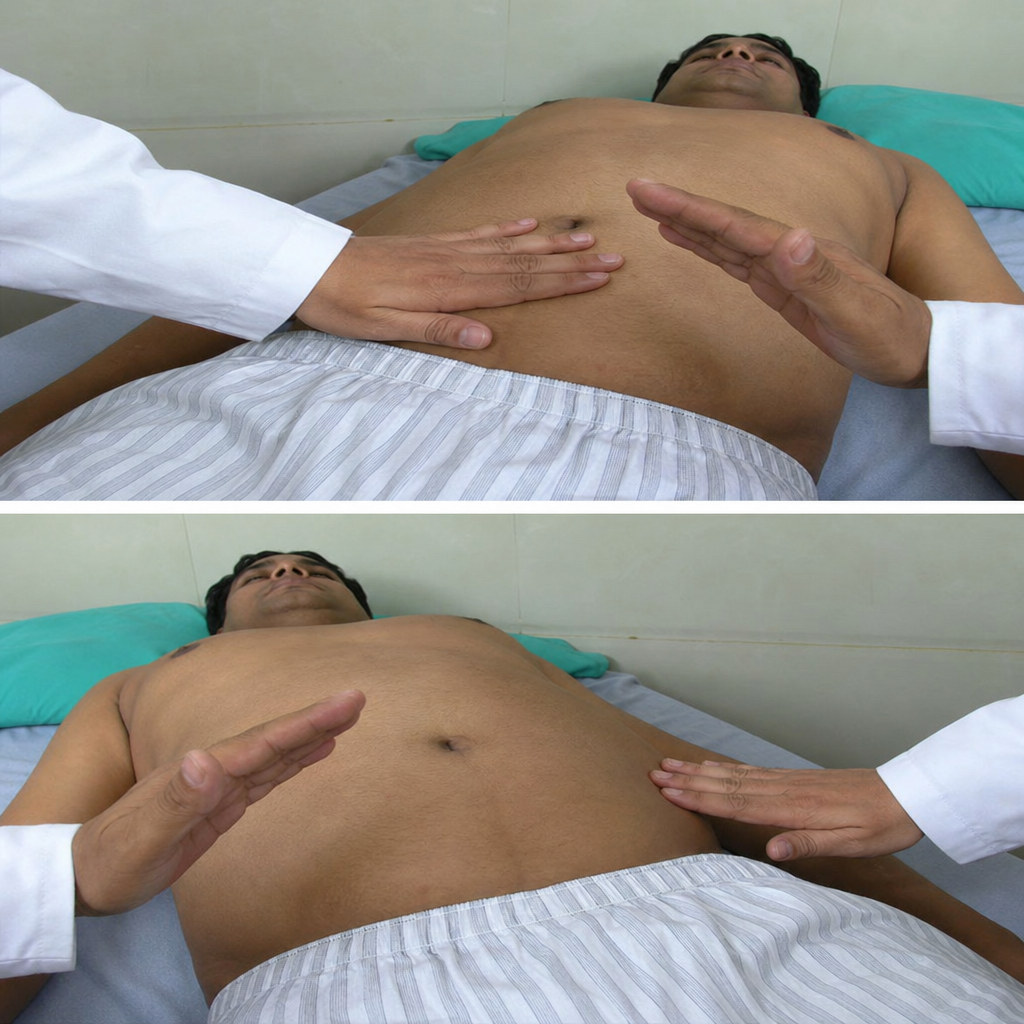
Which one of the following statements is TRUE regarding the clinical sign being elicited?
Which of the following statements is most appropriate?
Which of the following is a non-metabolic cause of abdominal pain?
Flapping tremors occur in all EXCEPT:
Practice by Chapter
Approach to Common Symptoms (Fever, Pain, Fatigue)
Practice Questions
Constitutional Symptoms and Their Differential Diagnosis
Practice Questions
Syncope and Presyncope
Practice Questions
Dizziness and Vertigo
Practice Questions
Dyspnea and Respiratory Distress
Practice Questions
Chest Pain Evaluation
Practice Questions
Abdominal Pain Assessment
Practice Questions
Headache Classification and Management
Practice Questions
Weight Loss and Cachexia
Practice Questions
Edema and Fluid Retention
Practice Questions
Want unlimited practice?
Get full access to all questions, explanations, and performance tracking.
Scan to download app