
Cardiology — MCQs

On this page
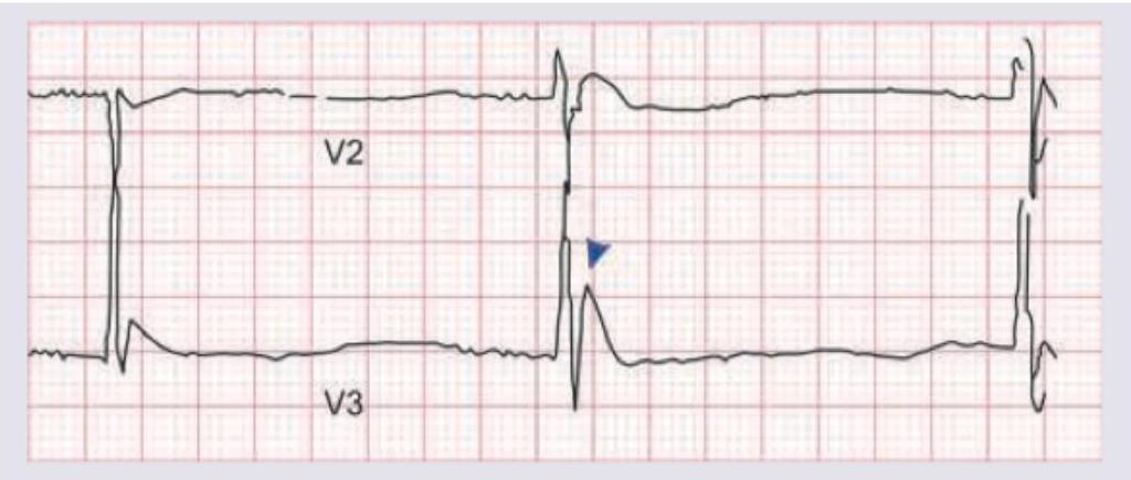
The following ECG finding is seen in all except:
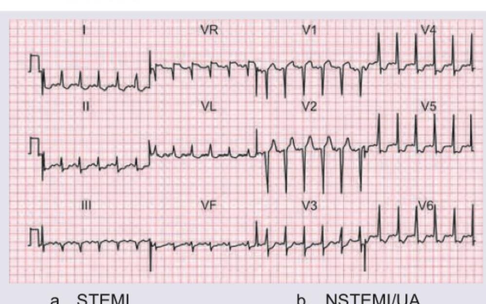
This ECG was recorded from a 65 -year-old man who had crescendo pattern episodes of chest pain. His current episode has been persisting at rest for 2.5 hours. Plasma troponin level was normal. Diagnosis is?
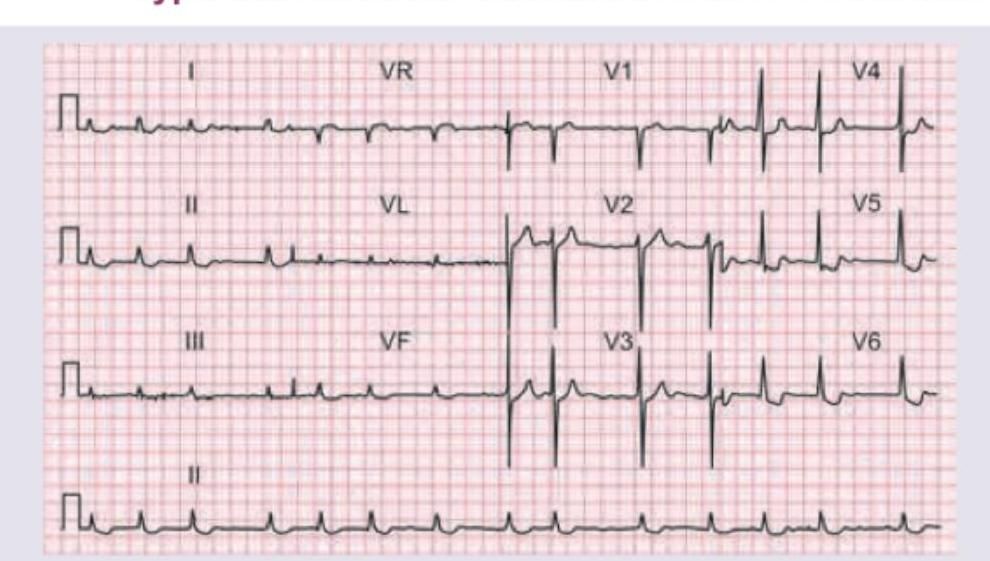
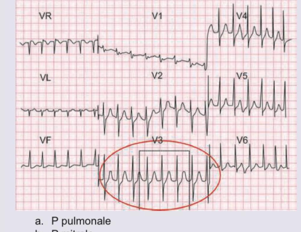
This ECG was recorded from a 60 -year-old woman with hypertension. Which is correct about the ECG shown?
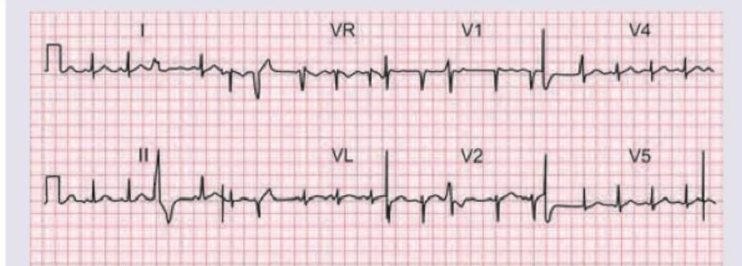
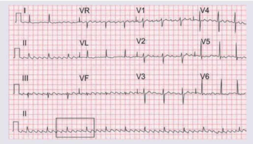
This ECG is of a 23 -year-old medical student who complained of an irregular heartbeat. Other than an irregular pulse, his heart was clinically normal. What does the ECG show?
In a patient with the given clinical sign which of the following ECG will be seen?

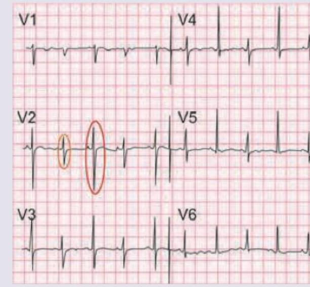
Which is correct about the ECG shown below?
A 25-year-old girl presents with history of recurrent syncopal attacks. Holter device was put and the recording is shown below. Which is correct about the recording?
A 30-year-old male was brought to the ER after a car crash. On admission, pulse is weak with BP=80 / 60 mm Hg. ECG is shown below. Right heart catheter is placed. Which is the most consistent value with patient's diagnosis?
The murmur of mitral regurgitation is best heard at
Which of the following statements is correct regarding the Opening Snap (OS) in a patient of mitral stenosis?
Practice by Chapter
Coronary Artery Disease and Angina
Practice Questions
Acute Coronary Syndromes
Practice Questions
Heart Failure
Practice Questions
Cardiac Arrhythmias
Practice Questions
Valvular Heart Diseases
Practice Questions
Cardiomyopathies
Practice Questions
Pericardial Diseases
Practice Questions
Congenital Heart Disease in Adults
Practice Questions
Hypertension and Hypertensive Emergencies
Practice Questions
Pulmonary Hypertension
Practice Questions
Non-invasive Cardiac Diagnostics
Practice Questions
Preventive Cardiology
Practice Questions
Want unlimited practice?
Get full access to all questions, explanations, and performance tracking.
Scan to download app