Which of the following is NOT seen in association with reverse splitting of S2?
A patient with angina, exertional syncope, and left ventricular hypertrophy is diagnosed with aortic stenosis. What is the predicted lifespan of this patient?
What is the most common cause of mitral valve disease?
Unequal pulses in upper and lower extremities (i.e., radio-femoral delay) is/are seen in which of the following conditions?
A 70-year-old male patient presented to the emergency department with pain in the epigastrium and difficulty in breathing for 6 hours. On examination, his heart rate was 56 beats per minute and the blood pressure was 106/60 mm Hg. Chest examination was normal. The patient has been taking omeprazole for gastroesophageal reflux disease for the last 6 months. What should be the initial investigation?
Which one of the following findings is NOT associated with left-sided heart failure?
Which of the following is NOT true regarding murmurs heard in hypertrophic obstructive cardiomyopathy?
Water hammer pulse is typically seen in which condition?
What is the most common cause of sudden cardiac death?
The Bundle of Kent is associated with which of the following conditions?
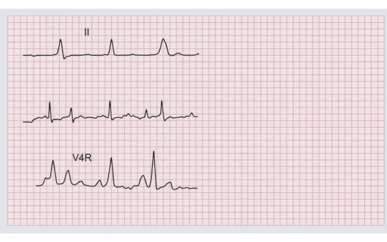
Explanation: **Explanation:** The second heart sound (S2) consists of two components: **A2 (Aortic)** and **P2 (Pulmonary)**. In a normal physiological state, A2 precedes P2. **Reverse (Paradoxical) splitting** occurs when there is a significant delay in the closure of the aortic valve, causing A2 to occur *after* P2. **Why Complete Heart Block (CHB) is the correct answer:** In Complete Heart Block, the atria and ventricles beat independently (AV dissociation) [1], [3]. While it can cause a "variable intensity" of S1 (Bruit de Canon), it does not inherently cause a consistent delay in left ventricular ejection that would lead to reverse splitting of S2. Therefore, it is not typically associated with this finding. **Analysis of Incorrect Options (Causes of Reverse Splitting):** * **LBBB (Option A):** This is the most common cause. Delayed electrical activation of the left ventricle leads to delayed mechanical contraction and late closure of the aortic valve. * **Systolic Hypertension (Option C):** High systemic pressure creates increased resistance to left ventricular ejection, prolonging the ejection time and delaying A2. * **Aortic Stenosis (Option D):** The narrowed valve orifice causes prolonged left ventricular ejection time to force blood through the obstruction, delaying A2 [4]. **NEET-PG High-Yield Pearls:** 1. **Paradoxical Split:** The split narrows during inspiration and widens during expiration (the opposite of physiological splitting). 2. **Wide Fixed Split:** Classically seen in **Atrial Septal Defect (ASD)** [2]. 3. **Wide Variable Split:** Seen in **RBBB** or Pulmonary Stenosis (where P2 is delayed). 4. **Soft/Absent A2:** A classic sign of severe calcific Aortic Stenosis [4].
Explanation: This question tests your knowledge of the natural history of symptomatic **Aortic Stenosis (AS)**. Once symptoms appear in a patient with severe AS, the prognosis declines sharply without surgical intervention (Aortic Valve Replacement). [2] ### **Explanation of the Correct Answer** The survival of patients with symptomatic aortic stenosis follows a classic timeline based on the presenting symptom, often remembered by the mnemonic **"A-S-D"**: 1. **A**ngina: Average survival is **5 years**. 2. **S**yncope: Average survival is **3 years**. 3. **D**yspnea (Heart Failure): Average survival is **2 **years**. In this clinical vignette, the patient presents with **exertional syncope**, which correlates with a predicted lifespan of **3 years**. Syncope in AS occurs due to the inability of the heart to increase cardiac output across a fixed orifice during exercise, leading to cerebral hypoperfusion. ### **Analysis of Incorrect Options** * **Option A (1 year):** While severe AS is life-threatening, the average survival for syncope is longer than one year. A 1-year survival is more characteristic of end-stage refractory heart failure. * **Option B (2 years):** This is the predicted survival for patients presenting with **Dyspnea/Heart Failure**, which carries the worst prognosis among the classic triad. * **Option D (4 years):** This does not correspond to any specific milestone in the classic AS survival timeline. Angina (the earliest symptom) has a 5-year survival. [1] ### **High-Yield Clinical Pearls for NEET-PG** * **Classic Triad:** Angina, Syncope, and Dyspnea (in order of worsening prognosis). * **Physical Exam:** Look for *Pulsus Parvus et Tardus* (slow-rising, low-volume pulse) and a mid-systolic ejection murmur that radiates to the carotids. [2] * **Reverse Splitting of S2:** Occurs because the aortic valve closes after the pulmonic valve due to prolonged LV ejection time. * **Management:** Medical therapy is generally ineffective; **Surgical Aortic Valve Replacement (SAVR)** or **TAVI** is the definitive treatment for symptomatic severe AS. [3]
Explanation: **Explanation:** **Rheumatic Heart Disease (RHD)**, resulting from **Rheumatic Fever**, remains the most common cause of acquired mitral valve disease worldwide, particularly in developing countries like India [1]. It is the leading cause of **Mitral Stenosis (MS)** and a frequent cause of Mitral Regurgitation (MR). The underlying mechanism involves an autoimmune response (molecular mimicry) following a Group A Streptococcal infection, leading to chronic inflammation, commissural fusion, and "fish-mouth" deformity of the valve. **Analysis of Incorrect Options:** * **Infective Endocarditis (A):** While it can cause acute, severe mitral regurgitation due to leaflet destruction or chordae rupture [2], it is less common than RHD and typically presents as an acute febrile illness rather than chronic valvular disease. * **Myxoma (B):** Atrial myxomas are the most common primary cardiac tumors. While they can physically obstruct the mitral orifice (mimicking mitral stenosis), they are rare clinical entities compared to the prevalence of RHD. * **Tuberculosis (C):** TB primarily affects the pericardium (causing constrictive pericarditis). It does not directly involve the endocardium or heart valves. **High-Yield Clinical Pearls for NEET-PG:** * **Mitral Stenosis:** RHD is the cause in nearly **99%** of cases of MS. * **Auscultation:** Look for a loud S1, an Opening Snap (OS), and a mid-diastolic rumbling murmur [3]. * **Mitral Regurgitation:** In developed nations, **Mitral Valve Prolapse (MVP/Myxomatous degeneration)** has overtaken RHD as the most common cause of isolated MR. * **Aschoff Bodies:** These are the pathognomonic histological features of acute rheumatic carditis.
Explanation: **Explanation:** The hallmark of **Coarctation of the Aorta (CoA)** is a significant pressure gradient across the narrowed segment, typically located just distal to the left subclavian artery (**Post-ductal**). This narrowing results in high blood pressure in the upper extremities and low blood pressure/delayed flow in the lower extremities, clinically manifesting as **radio-femoral delay** [1] and weak femoral pulses [2]. **Analysis of Options:** * **Post-ductal Coarctation (Correct):** This is the classic cause of radio-femoral delay [1]. The obstruction is distal to the origin of the brachiocephalic and left subclavian arteries, ensuring the upper body is well-perfused while the lower body receives blood via collaterals or the narrowed lumen. * **Aortic Dissection:** While it can cause *asymmetric* pulses (e.g., right vs. left arm), it typically presents with an acute, tearing chest pain. Radio-femoral delay is not its defining characteristic unless the dissection flap specifically occludes the distal aorta. * **Supra-valvular Aortic Stenosis:** This often presents with **differential pulses between the two arms** (Williams Syndrome). The jet of blood is directed toward the innominate artery (right arm), making the right radial pulse stronger than the left (the "reverse" of typical CoA). * **Sub-valvular Aortic Stenosis:** This is a fixed or dynamic obstruction (like HOCM) below the valve. It affects the entire systemic circulation equally, so no radio-femoral delay is observed. **High-Yield Clinical Pearls for NEET-PG:** * **Chest X-ray:** Look for the **"3" sign** (indentation of the aorta) and **rib notching** (due to dilated intercostal collateral arteries; usually involves 3rd to 8th ribs). * **Turner Syndrome:** 15-20% of patients with Turner Syndrome have Coarctation of the Aorta [1]. * **Associated Lesion:** Bicuspid aortic valve is the most common associated cardiac anomaly (up to 70% of cases). * **Physical Exam:** Always check for a systolic murmur loudest in the **left infrascapular area**.
Explanation: **Explanation:** The primary clinical challenge in this scenario is to differentiate between a benign gastrointestinal issue and a life-threatening cardiac event. In an elderly patient (70 years old) presenting with **epigastric pain** and **shortness of breath (dyspnea)**, the most critical diagnosis to rule out is an **Acute Coronary Syndrome (ACS)**, specifically an **Inferior Wall Myocardial Infarction (IWMI)** [1]. **Why ECG is the Correct Initial Investigation:** 1. **Atypical Presentation:** Elderly patients, females, and diabetics often present with "anginal equivalents" like epigastric pain or dyspnea rather than classic retrosternal chest pain [1]. 2. **Clinical Clues:** The presence of **bradycardia (HR 56 bpm)** in the setting of epigastric pain is a classic sign of IWMI due to increased vagal tone or ischemia of the SA/AV node [1]. 3. **Rule of Priority:** In emergency medicine, "life before limb/organ" applies. An ECG is rapid, non-invasive, and can diagnose a STEMI immediately, allowing for time-sensitive reperfusion therapy [2], [3]. **Why Other Options are Incorrect:** * **Upper GI Endoscopy:** While the patient has a history of GERD, an endoscopy is invasive and contraindicated in an unstable patient with a potential acute MI. * **Ultrasound Abdomen:** Useful for cholecystitis or pancreatitis, but these are secondary differentials that should only be explored after ruling out cardiac causes. * **X-ray Chest:** While it can help rule out pneumonia or pneumothorax, it does not diagnose the most lethal possibility (MI) suggested by the bradycardia. **NEET-PG High-Yield Pearls:** * **Inferior Wall MI** often presents with epigastric pain, nausea, and bradycardia (Vagal stimulation) [1], [2]. * **The "Golden Hour":** Any patient above 40 years with upper abdominal pain must receive an ECG within 10 minutes of arrival to the ED. * **Omeprazole Trap:** Do not be distracted by the history of GERD; "masking" of cardiac pain by antacids is a common clinical pitfall.
Explanation: ### Explanation **1. Why Hepatojugular Reflux (HJR) is the Correct Answer:** Hepatojugular reflux is a clinical sign of **Right-Sided Heart Failure (RHF)**. It occurs when manual pressure over the liver increases venous return to a failing right ventricle that cannot compensate, leading to a sustained rise in the Jugular Venous Pressure (JVP). While chronic left-sided heart failure (LHF) can eventually lead to RHF (the most common cause), HJR specifically reflects right ventricular dysfunction or systemic venous congestion, not isolated LHF [2]. **2. Analysis of Incorrect Options (Findings associated with LHF):** * **Pulmonary Edema (Option A):** In LHF, the left ventricle fails to pump blood forward, causing increased pressure in the left atrium and pulmonary veins. This hydrostatic pressure forces fluid into the alveoli, leading to pulmonary edema [1]. * **Paroxysmal Nocturnal Dyspnea (Option C):** This is a classic symptom of LHF. When a patient lies flat, interstitial fluid from the lower extremities is redistributed into the central circulation. A failing left ventricle cannot handle this increased preload, resulting in acute pulmonary congestion at night [2]. * **Prerenal Azotemia (Option D):** LHF causes a "forward failure" state with decreased cardiac output. This leads to reduced renal perfusion, triggering an increase in Blood Urea Nitrogen (BUN) and Creatinine, known as prerenal azotemia [3]. **3. Clinical Pearls for NEET-PG:** * **Most common cause of RHF:** Left-sided heart failure [2]. * **Earliest symptom of LHF:** Exertional dyspnea [2]. * **Specific sign of LHF:** Pulsus alternans (alternating strong and weak peripheral pulses). * **HJR Technique:** Apply firm pressure over the RUQ for 10–30 seconds; a positive result is a sustained rise in JVP >3 cm. It is highly suggestive of an elevated Pulmonary Capillary Wedge Pressure (PCWP).
Explanation: In Hypertrophic Obstructive Cardiomyopathy (HOCM), the murmur is primarily caused by dynamic Left Ventricular Outflow Tract (LVOT) obstruction. The intensity of the murmur depends on the **Left Ventricular (LV) volume**: anything that decreases LV volume (decreased preload or afterload) increases the obstruction and the murmur, while anything that increases LV volume (increased preload or afterload) decreases the murmur. **Explanation of Options:** * **Option C (Correct):** Handgrip exercise increases **afterload** (systemic vascular resistance) [1]. Increased afterload pushes back against the LV, increasing the ventricular volume and "forcing" the outflow tract open [1]. This **decreases** the intensity of the HOCM murmur [2]. Therefore, the statement that it increases is false. * **Option A:** Squatting increases both **preload** (venous return) and **afterload**. This increases LV volume, which reduces the obstruction and **decreases** the murmur [2]. * **Option B:** The Valsalva maneuver (strain phase) decreases venous return (**preload**). This reduces LV volume, allowing the hypertrophied septum and mitral valve to appose more easily, thereby **increasing** the murmur. * **Option D:** Beta-blockers are negative inotropes. They decrease the force of contraction and increase diastolic filling time, which reduces the dynamic obstruction and **decreases** the murmur. **High-Yield Clinical Pearls for NEET-PG:** 1. **The "Rule of Two":** HOCM and Mitral Valve Prolapse (MVP) are the only two murmurs that **increase** with Valsalva and standing (decreased preload). 2. **Dynamic Nature:** Unlike Valvular Aortic Stenosis (which decreases with Valsalva) [3], the HOCM murmur changes significantly with bedside maneuvers. 3. **Handgrip Differentiation:** Handgrip increases the murmur of Mitral Regurgitation and VSD but decreases the murmur of HOCM and Aortic Stenosis.
Explanation: **Explanation:** **1. Why Aortic Regurgitation (AR) is correct:** The **Water Hammer pulse** (also known as Corrigan’s pulse or collapsing pulse) is a hallmark of Aortic Regurgitation [1]. It is characterized by a rapid, forceful upstroke followed by a sudden, quick collapse. * **The Mechanism:** In AR, a large volume of blood is ejected into the aorta during systole (increased stroke volume), causing the sharp upstroke [1]. During diastole, blood rapidly flows backward into the left ventricle and forward into the peripheral circulation, leading to a precipitous drop in diastolic pressure. This results in a **wide pulse pressure**, which is the physiological basis for the "collapsing" sensation [1][2]. **2. Why other options are incorrect:** * **Aortic Stenosis (AS):** Characterized by **Pulsus Parvus et Tardus** (small volume and delayed peak) due to the obstructed outflow from the left ventricle. * **Aortic Stenosis and Regurgitation:** While both may coexist, the presence of AS often "dampens" the collapsing nature of the pulse. This combination typically results in **Pulsus Bisferiens** (a double-peaked systolic pulse). * **Mitral Regurgitation:** Usually presents with a normal or slightly reduced pulse volume [3]; it does not cause the wide pulse pressure necessary for a water hammer pulse. **3. High-Yield Clinical Pearls for NEET-PG:** * **Best way to elicit:** Palpate the radial pulse with the palm of your hand while elevating the patient's arm above their head (gravity accentuates the diastolic backflow). * **Associated Signs of AR:** * **de Musset’s sign:** Head nodding with each heartbeat [1]. * **Quincke’s sign:** Capillary pulsations in the nail bed. * **Traube’s sign:** "Pistol shot" sounds heard over the femoral artery. * **Duroziez’s sign:** Systolic and diastolic murmurs heard over the femoral artery when compressed. * **Differential Diagnosis:** Other high-output states like Patent Ductus Arteriosus (PDA), Arteriovenous fistulas, and severe anemia can also cause a collapsing pulse.
Explanation: ### Explanation **1. Why Ventricular Fibrillation (VF) is Correct:** Sudden Cardiac Death (SCD) is most commonly triggered by a lethal cardiac arrhythmia. Among these, **Ventricular Fibrillation (VF)** is the most frequent terminal rhythm, accounting for approximately 75–80% of cases [2]. In VF, the ventricles quiver rapidly and irregularly instead of pumping blood [1], leading to an immediate loss of cardiac output, circulatory collapse, and death within minutes if not defibrillated [3]. The most common underlying structural cause leading to VF is **Coronary Artery Disease (CAD)**, particularly acute myocardial ischemia or scarring from a previous infarct. **2. Why the Other Options are Incorrect:** * **Atrial Fibrillation (AF):** While AF is the most common sustained arrhythmia, it involves the atria. It increases the risk of stroke (embolic events) and heart failure but is rarely a direct cause of sudden death as the AV node protects the ventricles from the rapid atrial rate. * **PSVT:** These are typically narrow-complex tachycardias (like AVNRT). While they cause palpitations, dizziness, or syncope, they are almost never life-threatening in patients with normal heart structures. * **A-V Block:** While complete heart block (3rd degree) can cause asystole or severe bradycardia leading to death, it is a much less common cause of SCD compared to tachyarrhythmias like VF. **3. High-Yield Clinical Pearls for NEET-PG:** * **Most common underlying pathology for SCD:** Coronary Artery Disease (CAD). * **Most common cause of SCD in young athletes (<35 years):** Hypertrophic Cardiomyopathy (HCM). * **The "Golden Hour":** Survival rates for VF decrease by 7–10% for every minute that passes without defibrillation [2]. * **Primary Prevention:** In patients with low Ejection Fraction (<35%), an Implantable Cardioverter Defibrillator (ICD) is the treatment of choice to prevent SCD.
Explanation: **Wolff-Parkinson-White (WPW) syndrome** is the correct answer because it is characterized by the presence of an accessory pathway known as the **Bundle of Kent** [1]. In a normal heart, the AV node is the only electrical bridge between the atria and ventricles. In WPW, the Bundle of Kent bypasses the AV node, leading to **pre-excitation** of the ventricles [2]. This results in the classic ECG triad: 1. **Short PR interval** (<0.12s) due to rapid conduction through the bypass tract [1]. 2. **Delta wave** (slurred upstroke of the QRS) representing early ventricular activation [1]. 3. **Widened QRS complex** (>0.12s) [1]. **Analysis of Incorrect Options:** * **Sick Sinus Syndrome:** Refers to SA node dysfunction leading to alternating bradycardia and tachycardia (Tachy-Brady syndrome); it does not involve accessory pathways. * **Lown-Ganong-Levine (LGL) Syndrome:** Associated with the **James fibers** (intranodal accessory pathway). It presents with a short PR interval but a **normal QRS** (no delta wave) [1]. * **Stokes-Adams Syndrome:** Refers to sudden syncope caused by a complete heart block or severe arrhythmia leading to decreased cardiac output. **High-Yield Clinical Pearls for NEET-PG:** * **Drug Contraindication:** Avoid **ABCD** (Atropine/Adenosine, Beta-blockers, Calcium channel blockers, Digoxin) in WPW with Atrial Fibrillation, as they block the AV node and may force conduction through the Bundle of Kent, risking Ventricular Fibrillation [2]. * **Treatment of Choice:** Radiofrequency ablation of the accessory pathway. * **Association:** WPW syndrome is sometimes associated with **Ebstein’s anomaly**.
Coronary Artery Disease and Angina
Practice Questions
Acute Coronary Syndromes
Practice Questions
Heart Failure
Practice Questions
Cardiac Arrhythmias
Practice Questions
Valvular Heart Diseases
Practice Questions
Cardiomyopathies
Practice Questions
Pericardial Diseases
Practice Questions
Congenital Heart Disease in Adults
Practice Questions
Hypertension and Hypertensive Emergencies
Practice Questions
Pulmonary Hypertension
Practice Questions
Non-invasive Cardiac Diagnostics
Practice Questions
Preventive Cardiology
Practice Questions
Get full access to all questions, explanations, and performance tracking.
Start For Free