Which of the following is NOT true regarding the initiation and titration of beta-blocker therapy in a patient with congestive heart failure?
All the following findings would be expected in a person with coarctation of the aorta except:
Lutembacher syndrome includes all except:
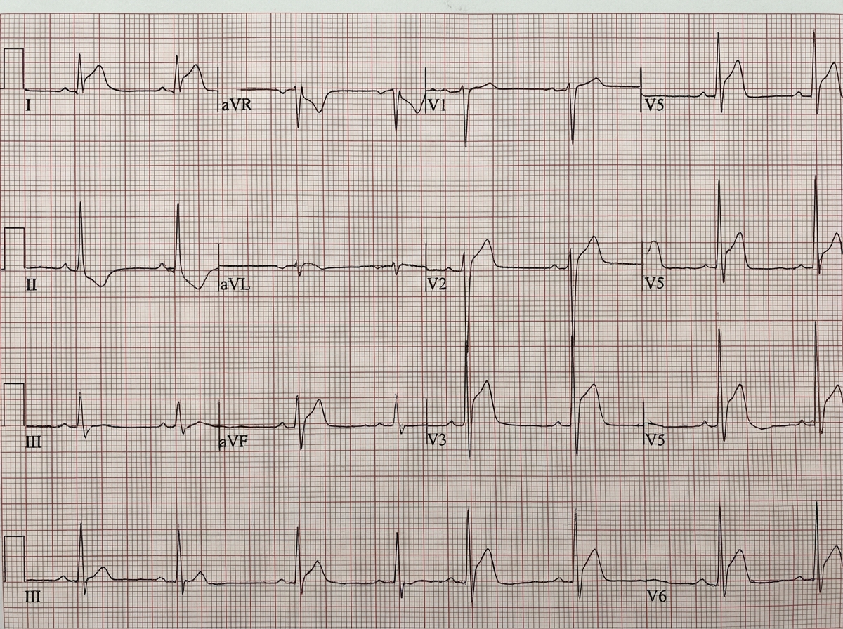
A 25-year-old patient with a history of recent respiratory tract infection complains of severe chest pain at rest. The ECG of the patient is given. What is the most probable diagnosis?

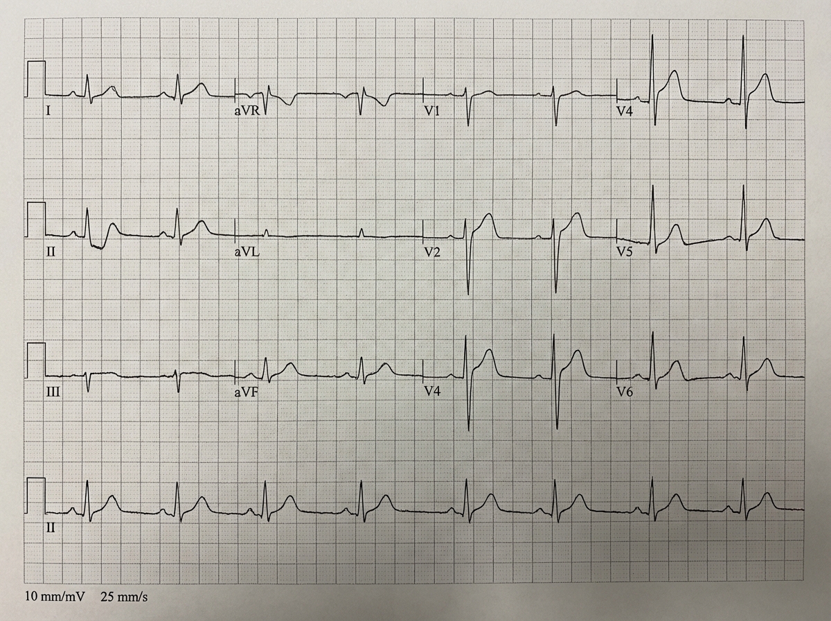
A young man reports retrosternal chest pain over the last few hours. The pain is not related to exertion and is relieved significantly on sitting up and leaning forward. What is the likely diagnosis?
A patient presents with low-grade fever. The ECG is shown below. What is the most probable diagnosis?

What is the drug of choice in patients with Wolff-Parkinson-White syndrome experiencing atrial fibrillation?
Which of the following statements regarding atrial fibrillation is false?
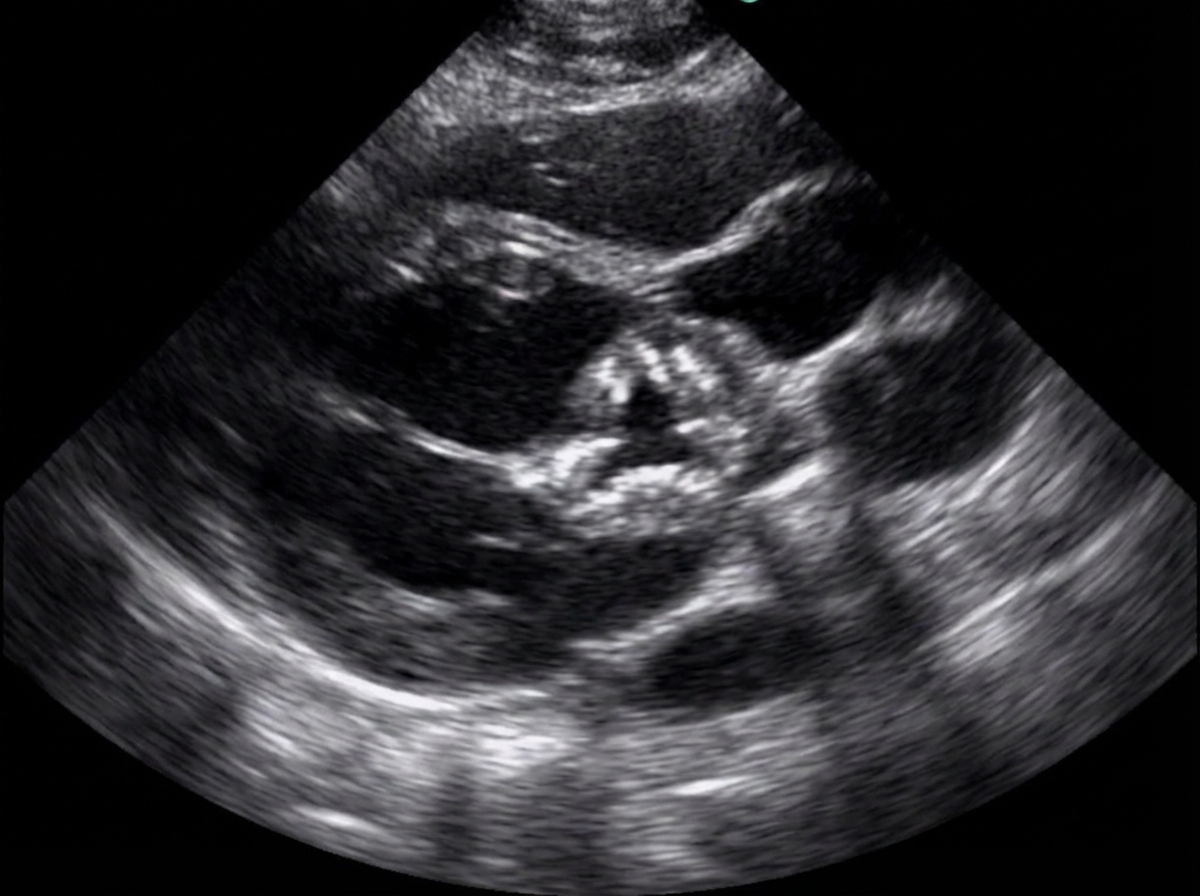
A 38-year-old woman presented with shortness of breath and fatigue. Her history is unremarkable except for a vague history of fever and joint pain as a child. She notes some recent fatigue and difficulty in sleeping that she attributes to job-related stress. On examination, her heart rate is 120 beats/min. Echocardiogram revealed the following finding. Auscultation of the heart indicates a harsh systolic murmur during left ventricular ejection. Which of the following pathological findings is not related with the underlying etiology?

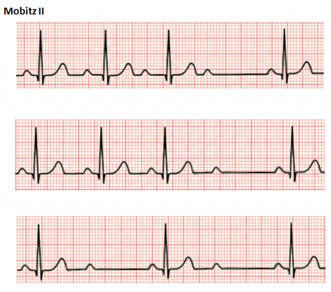
Comment on the diagnosis of the ECG tracing shown below?

Explanation: ### Explanation In the management of Congestive Heart Failure (CHF), the initiation of beta-blockers follows the mantra: **"Start low, go slow."** [1] **Why Option A is the Correct Answer (The "NOT True" statement):** Beta-blockers possess negative inotropic effects. If started at "optimum" (target) doses immediately, they can cause an acute drop in cardiac output, leading to a worsening of heart failure symptoms or pulmonary edema. Therefore, they must be initiated only when the patient is **hemodynamically stable and euvolemic**, starting at very low doses [3]. **Analysis of Other Options:** * **Option B:** Doses are typically doubled every 2–4 weeks as tolerated by the patient until the target dose (proven in clinical trials) or the maximum tolerated dose is reached [1]. * **Option C:** Patients in NYHA Class III and IV are at higher risk of decompensation. Initiation in these patients requires careful monitoring for fluid retention and bradycardia [2]. * **Option D:** Large-scale trials (like MERIT-HF, COPERNICUS, and CIBIS-II) have established **Carvedilol, Metoprolol succinate, and Bisoprolol** as the gold standard beta-blockers that reduce mortality in CHF [1]. **High-Yield Clinical Pearls for NEET-PG:** * **Contraindications:** Acute decompensated HF (cold/wet phase), symptomatic bradycardia, second or third-degree heart block, and severe reactive airway disease [3]. * **Carvedilol:** A non-selective beta-blocker with alpha-1 blocking properties, providing additional peripheral vasodilation [1]. * **Metoprolol:** Only the **succinate** (extended-release) form is evidence-based for mortality benefit in HF, not the tartrate form. * **Benefit:** Beta-blockers reduce "remodeling," decrease arrhythmias, and prevent the cardiotoxic effects of chronic catecholamine exposure [1].
Explanation: **Explanation:** **Coarctation of the Aorta (CoA)** is a localized narrowing of the aortic lumen, typically near the insertion of the ductus arteriosus. **Why Option C is the correct answer:** Patients with CoA generally maintain a **normal or even augmented cardiac output** during exercise [4]. While the narrowing increases afterload, the left ventricle undergoes compensatory hypertrophy to maintain stroke volume. During exercise, the heart responds to sympathetic stimulation by increasing heart rate and contractility [4]. Although exercise significantly exacerbates upper-body hypertension, it does not typically result in an "inability to augment" output unless there is advanced, end-stage heart failure. **Analysis of Incorrect Options:** * **Option A:** The systolic murmur is due to flow across the stenosis (heard best over the back). The **high-pitched diastolic murmur** is a classic finding representing an associated **Bicuspid Aortic Valve (BAV)**, which occurs in ~50-80% of CoA cases, leading to aortic regurgitation [2], [3]. * **Option B:** If the coarctation is **pre-subclavian** (proximal to the origin of the left subclavian artery), the blood pressure in the right arm will be significantly higher than in the left arm. * **Option D:** Persistent hypertension is a common long-term complication even after successful surgical repair. This is attributed to permanent changes in vascular compliance, altered baroreceptor sensitivity, and activation of the Renin-Angiotensin-Aldosterone System (RAAS). **NEET-PG High-Yield Pearls:** * **Classic Sign:** Radio-femoral delay and BP limb discrepancy (Upper limb > Lower limb) [1]. * **X-ray Findings:** **"3" sign** (indentation of the aorta) and **Rib notching** (due to collateral flow through dilated intercostal arteries; usually involves 3rd to 8th ribs). * **Association:** Strongly associated with **Turner Syndrome (45, XO)** [1]. * **Gold Standard Diagnosis:** CT Angiography or Cardiac MRI.
Explanation: Explanation: Lutembacher Syndrome is a specific clinical entity defined by the combination of acquired Mitral Stenosis (MS) and a congenital Atrial Septal Defect (ASD). 1. **Why Option C is correct:** Ventricular Septal Defect (VSD) is **not** a component of Lutembacher syndrome. The syndrome specifically involves an inter-atrial communication, not an inter-ventricular one. Therefore, VSD is the "except" in this list [1]. 2. **Why other options are incorrect:** * **Mitral Stenosis (Option A):** This is a core component. While usually acquired (rheumatic), it can rarely be congenital. * **Atrial Septal Defect (Option B):** This is the classic congenital component, most commonly of the *ostium secundum* type [1]. * **Left to Right Shunt (Option D):** In Lutembacher syndrome, the MS increases left atrial pressure, which significantly augments the left-to-right shunt across the ASD. This decompression of the left atrium actually delays the development of pulmonary venous congestion but leads to early right-sided heart failure. **High-Yield Clinical Pearls for NEET-PG:** * **Hemodynamics:** The ASD acts as a "decompression valve" for the left atrium. Consequently, the classic signs of MS (like loud S1 and opening snap) may be **diminished or absent** because the pressure gradient across the mitral valve is reduced. * **Clinical Presentation:** Patients typically present with features of right heart failure and pulmonary plethora rather than pulmonary edema. * **Auscultation:** You will primarily hear the signs of a large ASD (fixed split S2 and a mid-systolic flow murmur over the pulmonary area) [1]. * **Treatment:** The current treatment of choice is often percutaneous (PTMC for the mitral stenosis and device closure for the ASD).
Explanation: ***Acute pericarditis*** - **Recent respiratory tract infection** in a young patient followed by severe chest pain at rest is the classic presentation of **viral pericarditis**. - ECG typically shows **diffuse saddle-shaped ST elevation** with **PR depression** and no **reciprocal changes**, distinguishing it from myocardial infarction. *Acute myocardial infarction* - Would show **localized ST elevation** with **reciprocal ST depression** in opposite leads, not the diffuse pattern seen in pericarditis. - Uncommon in **young healthy patients** without significant risk factors like smoking, diabetes, or family history. *Prinzmetal angina* - Characterized by **transient ST elevation** during **coronary artery spasm**, typically occurring at night or early morning. - Pain is usually **brief and episodic**, not persistent severe chest pain, and resolves spontaneously with **nitroglycerin**. *Takotsubo cardiomyopathy* - Typically occurs in **postmenopausal women** following **emotional or physical stress**, not young patients with respiratory infections. - ECG shows **T-wave inversions** and **QT prolongation** rather than the ST elevation pattern of pericarditis.
Explanation: **Explanation:** The clinical presentation described is classic for **Acute Pericarditis**. The hallmark of pericardial pain is its **pleuritic nature** and its strong dependence on **body posture**. [3] 1. **Why Acute Pericarditis is correct:** The pain in pericarditis is caused by inflammation of the parietal pleura adjacent to the pericardium. Sitting up and leaning forward (the **"Tripod position"**) pulls the heart away from the diaphragm and adjacent pleura, thereby reducing friction and providing significant relief. [3] Conversely, the pain worsens in the supine position. 2. **Why other options are incorrect:** * **Rib Fracture:** Pain is typically localized, associated with a history of trauma, and worsens with direct palpation or deep inspiration, but is not specifically relieved by leaning forward. * **Angina Pectoris:** This is typically exertional, described as a "heaviness" or "pressure," and is relieved by rest or nitrates. [4] It does not change with posture. [2] * **Reflux Esophagitis:** While it causes retrosternal burning, it typically worsens after meals or when lying down, but it lacks the specific pleuritic/positional relief seen in pericarditis. **High-Yield Clinical Pearls for NEET-PG:** * **ECG Findings:** Look for **diffuse ST-segment elevation (concave upwards)** and **PR-segment depression** (the most specific early sign). [1] * **Physical Exam:** A **pericardial friction rub** (best heard with the diaphragm at the left lower sternal border) is pathognomonic. * **Treatment:** First-line treatment usually involves **NSAIDs (like Aspirin or Ibuprofen) plus Colchicine** to prevent recurrences. [1] * **Etiology:** Most common cause is viral (Coxsackievirus B). [1]
Explanation: ***Pericarditis*** - Classic ECG findings include **diffuse concave ST elevation** across multiple leads without reciprocal changes, often accompanied by **PR depression**. - The presence of **low-grade fever** is a typical clinical feature that supports the diagnosis of **acute pericarditis**. *Acute myocardial infarction* - ECG shows **convex ST elevation** in specific coronary territories with **reciprocal ST depression** in opposite leads. - Typically presents with **severe chest pain** and elevated **cardiac enzymes**, not the diffuse changes seen in pericarditis. *Old myocardial infarction* - ECG demonstrates **pathological Q waves** and **T wave inversions** in affected leads, indicating established **myocardial scarring**. - Would not present with **acute symptoms** like fever or the diffuse ST changes characteristic of pericarditis. *Hypothermia* - ECG classically shows **Osborn waves** (J waves) - positive deflections at the end of the QRS complex. - Patient would present with **low body temperature** and associated systemic signs, not fever as described in this case.
Explanation: ### Explanation In **Wolff-Parkinson-White (WPW) syndrome**, an accessory pathway (Bundle of Kent) bypasses the AV node [1]. When atrial fibrillation (AF) occurs in these patients, the accessory pathway can conduct impulses to the ventricles at extremely high rates because, unlike the AV node, it lacks a physiological delay (decremental conduction). **Why Procainamide is the Correct Answer:** Procainamide is a Class IA antiarrhythmic that **increases the refractory period of the accessory pathway**. By slowing conduction through the bypass tract, it reduces the ventricular rate and may even terminate the AF. In hemodynamically stable patients with WPW and AF, it is the drug of choice. (Note: If the patient is hemodynamically unstable, DC cardioversion is the immediate treatment). **Why the Other Options are Incorrect:** * **Digitalis (Digoxin), Verapamil, and Adenosine:** These are all **AV-nodal blocking agents**. By blocking the AV node, these drugs paradoxically favor conduction through the accessory pathway. This can lead to a rapid increase in ventricular rate, potentially degenerating into **Ventricular Fibrillation (VF)** and cardiac arrest. They are strictly contraindicated in WPW with AF. **Clinical Pearls for NEET-PG:** * **Classic Triad of WPW:** Short PR interval (<0.12s), Delta wave (slurred upstroke of QRS), and Wide QRS complex [1]. * **FBI Sign:** AF in WPW is often described as **F**ast, **B**road (wide QRS), and **I**rregular. * **Definitive Treatment:** Radiofrequency ablation of the accessory pathway. * **Avoid "ABCD" in WPW + AF:** **A**denosine, **B**eta-blockers, **C**alcium channel blockers (Verapamil/Diltiazem), and **D**igoxin.
Explanation: ### Explanation **Why Option A is the Correct (False) Statement:** In the management of Atrial Fibrillation (AF), **brain imaging (CT or MRI) is not routinely performed** for every patient [1]. It is only indicated if the patient presents with focal neurological deficits suggestive of an acute stroke or a Transient Ischemic Attack (TIA). Routine management focuses on rate/リズム control and thromboembolism prophylaxis based on risk scores, rather than preemptive neuroimaging [1]. **Analysis of Other Options:** * **Option B (P waves are absent):** This is a hallmark ECG finding. In AF, organized atrial depolarization is replaced by rapid, chaotic electrical activity (fibrillatory waves), leading to the absence of distinct P waves and an "irregularly irregular" ventricular rhythm [1]. * **Option C (Anticoagulants are typically added):** AF increases the risk of atrial thrombus formation (especially in the left atrial appendage) [1]. Anticoagulation (Warfarin or NOACs like Apixaban) is a cornerstone of therapy, guided by the **CHA₂DS₂-VASc score** to prevent embolic stroke [1]. * **Option D (Palpitations are common):** Palpitations are the most frequent presenting symptom due to the rapid and irregular heartbeat. Other symptoms include dyspnea, fatigue, and lightheadedness. **High-Yield Clinical Pearls for NEET-PG:** * **Most common chronic arrhythmia:** Atrial Fibrillation [1]. * **Investigation of Choice:** 12-lead ECG [1]. * **CHA₂DS₂-VASc Score:** Used to decide the need for anticoagulation (Score ≥2 in men or ≥3 in women generally requires anticoagulation) [1]. * **HAS-BLED Score:** Used to assess the 1-year risk of major bleeding in patients on anticoagulation. * **Treatment Strategy:** Hemodynamically unstable patients require immediate **synchronized cardioversion**. Stable patients are managed with rate control (Beta-blockers, CCBs, or Digoxin) or rhythm control [1].
Explanation: ***Pulmonary hypertension*** - **Pulmonary hypertension** typically results from elevated **left atrial pressure** due to **mitral stenosis**, not aortic stenosis. - **Aortic stenosis** primarily causes **left ventricular pressure overload** without directly affecting pulmonary circulation. *Aortic regurgitation* - **Rheumatic fever** commonly affects both **aortic** and **mitral valves**, causing mixed lesions including **aortic regurgitation**. - **Bicuspid aortic valve** from rheumatic disease can lead to both **stenosis** and **regurgitation** over time. *Left ventricular hypertrophy* - **Aortic stenosis** causes **pressure overload** on the left ventricle, leading to **concentric hypertrophy**. - **LVH** is a direct compensatory mechanism to maintain **cardiac output** against increased **afterload**. *Mitral stenosis* - **Rheumatic fever** classically causes **mitral stenosis** through **commissural fusion** and **leaflet thickening**. - **Mitral stenosis** is the most common **rheumatic valvular lesion** and often coexists with **aortic involvement**.
Explanation: ***Second degree AV block type 2, infranodal*** - Shows **constant PR intervals** with **sudden dropped QRS complexes** without preceding PR prolongation, characteristic of **Mobitz Type II**. - The **wide QRS complexes** and **infranodal location** (His-Purkinje system) indicate a more serious conduction defect requiring **pacemaker consideration**. *First degree AV block, intranodal* - Would show **prolonged PR interval >200ms** but **every P wave** would be followed by a QRS complex. - No **dropped beats** would be present, unlike the intermittent AV conduction seen here. *Second degree AV block type 1, intranodal* - Characterized by **progressive PR prolongation** followed by a dropped QRS (**Wenckebach phenomenon**). - Typically has **narrow QRS complexes** and occurs at the **AV nodal level**, not infranodal. *Third degree AV block* - Would show **complete AV dissociation** with **independent P waves and QRS complexes**. - The **ventricular rate** would be slower and **regular**, with no relationship between P waves and QRS complexes.
Coronary Artery Disease and Angina
Practice Questions
Acute Coronary Syndromes
Practice Questions
Heart Failure
Practice Questions
Cardiac Arrhythmias
Practice Questions
Valvular Heart Diseases
Practice Questions
Cardiomyopathies
Practice Questions
Pericardial Diseases
Practice Questions
Congenital Heart Disease in Adults
Practice Questions
Hypertension and Hypertensive Emergencies
Practice Questions
Pulmonary Hypertension
Practice Questions
Non-invasive Cardiac Diagnostics
Practice Questions
Preventive Cardiology
Practice Questions
Get full access to all questions, explanations, and performance tracking.
Start For Free