
Cardiology — MCQs

On this page
A 55-year-old female presents with Levine sign, hiccups, and vomiting episodes. On examination, HR=50/min with BP =100/60 mm Hg with elevated JVP. ECG technician is yet to arrive. Which coronary artery is likely to be involved?
Congenital long QT syndrome can lead to which of the following?
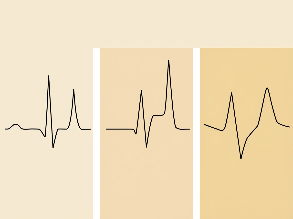
Which condition is most likely associated with the given ECG tracing?
What is the most common valvular lesion seen with carcinoid syndrome?
Wolff-Parkinson-White (WPW) syndrome is characterized by which of the following electrocardiographic findings?
Which one of the following is of most serious prognostic significance in a patient of essential hypertension?
Increased AV nodal blockade leads to termination of tachycardia in all except?
A 48-year-old, previously healthy woman reports having suddenly lost consciousness four times in the past 6 months. In three instances, she was unconscious for only a few minutes. After the fourth episode 1 month ago, she was unconscious for 6 hours and had weakness in her right arm and difficulty speaking. On physical examination, she is afebrile, and her blood pressure is normal. No murmurs are auscultated. She has good carotid pulses with no bruits. Which of the following cardiac lesions is most likely to be present in this woman?
A 25-year-old male diagnosed with Atrial Septal Defect (ASD) presents with a murmur similar to Mitral Regurgitation (MR) on examination and an ECG showing left axis deviation of 40 degrees. What is the most likely underlying condition?
What is a main disadvantage of stenting?
Practice by Chapter
Coronary Artery Disease and Angina
Practice Questions
Acute Coronary Syndromes
Practice Questions
Heart Failure
Practice Questions
Cardiac Arrhythmias
Practice Questions
Valvular Heart Diseases
Practice Questions
Cardiomyopathies
Practice Questions
Pericardial Diseases
Practice Questions
Congenital Heart Disease in Adults
Practice Questions
Hypertension and Hypertensive Emergencies
Practice Questions
Pulmonary Hypertension
Practice Questions
Non-invasive Cardiac Diagnostics
Practice Questions
Preventive Cardiology
Practice Questions
Want unlimited practice?
Get full access to all questions, explanations, and performance tracking.
Scan to download app