Which type of cardiomyopathy is associated with alcohol abuse?
A 30-year-old man presents with cramping gluteal pain after walking 500m. Which vascular condition is most likely involved?
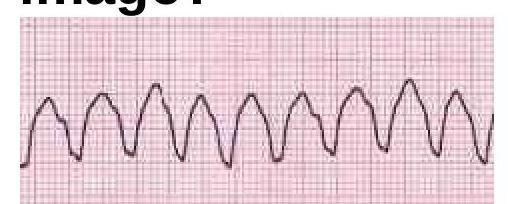
Osborn J waves are seen in which of the following conditions?
What electrolyte imbalance is most commonly associated with prominent U waves on ECG?
Roth spots are associated with which of the following conditions?
In which of the following conditions is the implantation of an Automatic Implantable Cardioverter Defibrillator (AICD) indicated?
In which condition does the Ankle-Brachial Pressure Index (ABPI) increase artificially?
What are the potential causes of cardiogenic shock excluding myocardial infarction?
What does the CEAP score classify?
Which drug is used as an adjunct to epinephrine in refractory ventricular fibrillation/ventricular tachycardia during cardiac arrest?
Explanation: ### Dilated cardiomyopathy - Chronic **alcohol abuse** is a well-established cause of **dilated cardiomyopathy**, leading to weakening and enlargement of the ventricles [1]. - This condition results in impaired systolic function and can cause **heart failure** [1]. *Hypertrophic cardiomyopathy* - This condition is primarily characterized by **pathological thickening of the heart muscle**, often genetic, and is not directly caused by alcohol abuse. - It leads to issues with relaxation and filling of the heart, rather than dilation and weakness. *Pericarditis* - **Pericarditis** is the inflammation of the sac surrounding the heart (pericardium), most commonly caused by viral infections, autoimmune diseases, or injury. - It is not directly linked to alcohol abuse as a primary cause. *Myocarditis* - **Myocarditis** is the inflammation of the heart muscle, often triggered by viral infections, autoimmune reactions, or certain medications. - While heavy alcohol use can weaken the heart, myocarditis is primarily an inflammatory process not directly caused by alcohol.
Explanation: ***Aortoiliac arterial disease*** - **Cramping gluteal pain** (claudication) is characteristic of **aortoiliac disease** due to insufficient blood flow to the gluteal muscles [1]. - The disease typically involves the **aorta** and/or the **iliac arteries**, which supply blood to the pelvis and lower limbs [1]. *Superficial femoral artery disease* - This condition primarily causes claudication in the **calf muscles**, not typically the gluteal region [1]. - The pain would be felt lower down the leg, corresponding to the muscle groups supplied by the **superficial femoral artery** [1]. *Popliteal artery disease* - **Popliteal artery disease** causes claudication in the **calf and foot** due to reduced blood flow to these distal areas [1]. - It would not typically present with gluteal pain, as the popliteal artery is located behind the knee. *No vascular involvement* - The presence of **cramping pain after exertion**, specifically claudication, strongly indicates significant vascular compromise [1]. - This symptom pattern is classic for **peripheral arterial disease**, ruling out no vascular involvement [1].
Explanation: ***Hypothermia*** - **Osborn J waves**, or **J waves**, are characteristic electrocardiogram (ECG) findings in patients with **hypothermia**. - These waves are small, positive deflections seen at the **junction of the QRS complex and the ST segment**, most prominent in the precordial leads. *Hypocalcemia* - Hypocalcemia typically manifests on an ECG as a **prolonged QT interval** due to delayed repolarization, which can increase the risk of arrhythmias. - It does not cause Osborn J waves. *Hyperkalemia* - Hyperkalemia causes distinct ECG changes including **tall, peaked T waves**, prolonged PR interval, widened QRS complex, and ultimately sine wave formation [1]. - Osborn J waves are not associated with hyperkalemia. *Hypokalemia* - Hypokalemia is associated with ECG changes such as **flattening or inversion of T waves**, prominent U waves, and ST-segment depression [1]. - It does not lead to the formation of Osborn J waves.
Explanation: ***Hypokalemia*** - **Prominent U waves** on an ECG are a classic sign of **hypokalemia**, indicating delayed repolarization of ventricular myocardial cells. - Other ECG changes associated with hypokalemia include **ST-segment depression**, **flattened T waves**, and a prolonged QT interval. *Hyponatremia* - **Hyponatremia** primarily affects neurological function and can lead to symptoms like confusion and seizures [1]. - It does not typically cause specific characteristic ECG changes such as **prominent U waves**. *Hypocalcemia* - **Hypocalcemia** is often associated with a **prolonged QT interval** on an ECG, which reflects delayed ventricular repolarization [2]. - However, it is not primarily linked to the appearance of prominent U waves. *Hypercalcemia* - **Hypercalcemia** is known to cause a **shortened QT interval** on the ECG due to accelerated repolarization. - It does not typically manifest with prominent U waves.
Explanation: **Acute leukaemia** - **Roth spots** are commonly observed in patients with **leukaemia**, characterized by **retinal hemorrhages** with pale centers due to leukemic cell infiltrates [1]. - The presence of these lesions is often an indicator of severe systemic disease [1]. *Bacterial endocarditis* - While **bacterial endocarditis** can cause various **ophthalmologic manifestations** like splinter hemorrhages and Janeway lesions, **Roth spots** are distinctly identified as hemorrhages with pale centers, which, though they can occur, are less characteristically associated than with leukaemia. - These hemorrhages in endocarditis are usually attributed to **immune complex deposition** or **septic emboli**. *Severe Hypertension* - **Severe hypertension** can lead to **hypertensive retinopathy**, presenting as **retinal hemorrhages**, cotton wool spots, and papilledema. - However, the classic description of **Roth spots** with their pale centers is not a direct or prominent feature of hypertensive retinopathy. *None of the options* - This option is incorrect because **Acute leukaemia** is clearly associated with **Roth spots**.
Explanation: ***Ventricular tachycardia with structural heart disease*** - An **AICD** is strongly indicated for patients with **sustained ventricular tachycardia (VT)** in the presence of **structural heart disease** due to the high risk of sudden cardiac death [1]. - In these cases, the AICD can deliver **therapy (antitachycardia pacing or defibrillation)** to terminate life-threatening arrhythmias [1]. *Syncope due to arrhythmias* - While syncope due to arrhythmias can be serious, an **AICD** is not always the first or only treatment and its indication depends on the specific arrhythmia and underlying cause. - Other treatments like **ablation**, **antiarrhythmic medications**, or a **pacemaker** might be more appropriate depending on the type of arrhythmia (e.g., bradycardia). *None of the options* - This option is incorrect because **ventricular tachycardia with structural heart disease** is a clear and well-established indication for AICD implantation [1]. - AICDs are a cornerstone in the secondary prevention of sudden cardiac death in high-risk patients. *Brugada syndrome* - While **Brugada syndrome** carries a risk of sudden cardiac death, AICD implantation is typically reserved for patients who have experienced **symptomatic arrhythmias** (e.g., syncope, aborted sudden cardiac death) or have certain high-risk features, not for all asymptomatic cases. - Risk stratification in Brugada syndrome is complex, and an AICD is not universally indicated for every diagnosed individual.
Explanation: ***Arteriosclerosis calcified arteries*** - **Arterial calcification** makes the vessels rigid and **non-compressible**, leading to falsely elevated blood pressure readings in the ankle. - This results in an **artificially high ABPI** (>1.3), masking the true extent of peripheral artery disease or even suggesting absence of disease [2]. *Ischemic ulcers* - **Ischemic ulcers** are a consequence of severe peripheral artery disease, which typically causes a **decreased ABPI** (<0.9) [2]. - The reduced blood flow cannot artificially increase the ABPI; rather, it indicates severe circulatory compromise. *Intermittent claudication* - **Intermittent claudication** is a symptom of peripheral artery disease, where the ABPI is usually **reduced during exercise** or **at rest** (<0.9) [1]. - This condition is characterized by arterial narrowing, not by stiffening that artificially inflates pressure readings. *DVT* - **Deep vein thrombosis (DVT)** primarily affects the venous system and **does not directly influence arterial pressure** measurements used for ABPI [2]. - While DVT can cause leg pain and swelling, it does not cause an artificial increase in arterial pressure readings in the ankle.
Explanation: ***acute mitral regurgitation*** - Severe acute mitral regurgitation can lead to **cardiogenic shock** by causing a sudden increase in left atrial pressure and pulmonary edema, reducing effective forward flow [1]. - This condition can result from **papillary muscle rupture** or dysfunction, often due to ischemia even in the absence of a large MI, or from infective endocarditis [1]. *ventricular septal rupture* - A ventricular septal rupture (VSR) is a mechanical complication of **myocardial infarction (MI)**, which is explicitly excluded in the question. - VSR creates a **left-to-right shunt**, leading to pulmonary overload and reduced systemic cardiac output, but it's fundamentally post-MI. *right heart failure* - While right heart failure can cause circulatory collapse, it is typically a component of **biventricular failure** or isolated in cases like severe pulmonary embolism [2]. - The question asks for causes of cardiogenic shock excluding MI, and pure **right heart failure** as a sole cause of global cardiogenic shock (affecting systemic perfusion) is less common without significant left heart involvement or severe pulmonary vascular disease. *All of the options* - This option is incorrect because **ventricular septal rupture** is almost exclusively a complication of myocardial infarction, which the question explicitly excludes. - Therefore, not all listed conditions are potential causes of cardiogenic shock *excluding MI*.
Explanation: ***Chronic venous disorder*** - The **CEAP classification** is a standardized system for classifying **chronic venous disorders** [1]. - It stands for **Clinical, Etiological, Anatomical, and Pathophysiological**, providing a comprehensive description of the disease. *Atrial disorders* - **Atrial disorders** refer to conditions affecting the atria of the heart, such as atrial fibrillation, and are classified using different systems. - The CEAP score has **no relevance** to cardiac conditions. *Neurological disorders* - **Neurological disorders** pertain to conditions of the nervous system and are classified by systems tailored to their specific characteristics. - The CEAP score is **not applicable** to neurological diagnoses. *Traumatic disorders* - **Traumatic disorders** result from physical injury and are classified based on the nature and severity of the injury. - The CEAP score is **irrelevant** for assessing or classifying traumatic injuries.
Explanation: ***Amiodarone infusion*** - **Amiodarone** is a **Class III antiarrhythmic** drug commonly used in advanced cardiac life support (ACLS) protocols for refractory **ventricular fibrillation (VF)** or **pulseless ventricular tachycardia (VT)** that persists despite defibrillation and epinephrine [1]. - It works by blocking potassium channels, prolonging repolarization and the refractory period, which helps to stabilize the electrical activity of the heart. *Atropine* - **Atropine** is an anticholinergic drug primarily used to treat **symptomatic bradycardia** by increasing heart rate. - It is not indicated for the treatment of **ventricular fibrillation** or **ventricular tachycardia** during cardiac arrest. *High dose vasopressin* - **Vasopressin** was previously included in some ACLS algorithms as an alternative to epinephrine for **vasoconstrictive effects**, but recent guidelines do not support its routine use in cardiac arrest. - While it can cause **vasoconstriction**, there is no evidence that high-dose vasopressin improves outcomes in refractory VF/VT over epinephrine. *Adenosine* - **Adenosine** is an antiarrhythmic drug used to treat **supraventricular tachycardias (SVTs)** by transiently blocking the AV node. - It is not effective for **ventricular fibrillation** or **ventricular tachycardia** and can even be harmful in these rhythms.
Coronary Artery Disease and Angina
Practice Questions
Acute Coronary Syndromes
Practice Questions
Heart Failure
Practice Questions
Cardiac Arrhythmias
Practice Questions
Valvular Heart Diseases
Practice Questions
Cardiomyopathies
Practice Questions
Pericardial Diseases
Practice Questions
Congenital Heart Disease in Adults
Practice Questions
Hypertension and Hypertensive Emergencies
Practice Questions
Pulmonary Hypertension
Practice Questions
Non-invasive Cardiac Diagnostics
Practice Questions
Preventive Cardiology
Practice Questions
Get full access to all questions, explanations, and performance tracking.
Start For Free