Erb's Point in cardiology refers to:
Turner syndrome is maximally associated with ?
Which of the following is the MOST common complication of untreated hypertension?
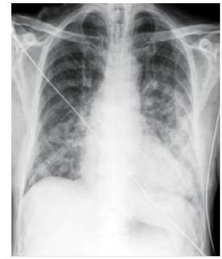
A hypertensive patient who is non-compliant with medication presents to you with sudden onset breathlessness. A chest x-ray was done, which is shown below. How will you manage this patient?

What is the normal range for the QRS axis in degrees?
Identify the cardiac condition represented in the image.

What is the most appropriate immediate management for a hemodynamically unstable patient with supraventricular tachycardia (SVT)?
Vegetation in mitral valve seen in which condition
Which of the following statements is true about the Bundle of Kent?
A patient presents to you with an irregularly irregular pulse of 120/minutes and a pulse deficit of 20. Which of the following would be the jugular venous pressure (JVP) finding?

Explanation: ***Left sternal border at 3rd intercostal space*** - **Erb's point** is a key auscultation site located at the **third intercostal space** along the **left sternal border**. - This location is particularly important because sounds from the **aortic** and **pulmonic valves** can often be heard here, as well as murmurs like the murmur of **aortic regurgitation**. *Right 2nd intercostal space* - The **right second intercostal space** at the sternal border is the primary auscultation site for the **aortic valve**. - While aortic sounds are heard at Erb's point, this specific location is not referred to as Erb's point. *Left 2nd intercostal space* - The **left second intercostal space** at the sternal border is the primary auscultation site for the **pulmonic valve**. - It is used to best hear sounds related to the pulmonary circulation, rather than being Erb's point. *Right 3rd intercostal space* - The **right third intercostal space** at the sternal border is not a primary or named auscultation point in the standard set of cardiology landmarks. - Auscultation in this area would primarily involve sounds transmitted from adjacent valve areas.
Explanation: **Coarctation of aorta** - **Coarctation of the aorta** is the most common cardiovascular abnormality associated with **Turner syndrome**, occurring in 5-20% of affected individuals [1]. - It is a **narrowing of the aorta**, typically near the ductus arteriosus, which can lead to hypertension and other cardiovascular complications [1]. *Horseshoe kidney* - While **renal anomalies** are common in Turner syndrome (occurring in 30-60%), **horseshoe kidney** is not the single most prevalent or *maximally associated* malformation. - Other renal abnormalities like duplicated collecting systems and pelvic kidneys are also frequently observed. *VSD* - **Ventricular septal defect (VSD)** is a type of congenital heart defect, but it is not as frequently associated with Turner syndrome as coarctation of the aorta [2]. - VSDs are among the most common congenital heart defects overall but are not the leading cardiovascular anomaly specific to Turner syndrome [3]. *ASD* - **Atrial septal defect (ASD)** is another congenital heart anomaly, but it is not the *maximally associated* cardiovascular defect in Turner syndrome. - Coarctation of the aorta and bicuspid aortic valve are more characteristic cardiovascular findings in this condition [1].
Explanation: ***Stroke*** - **Untreated hypertension** is a major risk factor for both **ischemic** and **hemorrhagic strokes**, damaging blood vessels in the brain [1]. - Sustained high blood pressure can lead to **atherosclerosis** and weakened blood vessel walls, increasing the likelihood of a stroke [1]. *Heart failure* - While hypertension is a leading cause of **heart failure**, it typically develops over a longer period as the heart works harder against elevated pressure, leading to remodeling and dysfunction [2]. - In terms of acute, severe complications, a stroke is often considered more immediately catastrophic. *Kidney damage* - **Hypertensive nephropathy** is a significant long-term complication where high blood pressure damages the small blood vessels in the kidneys, impairing their function. - This process is gradual and contributes to **chronic kidney disease**, but stroke often emerges as a more common and acute serious event. *Vision loss* - **Hypertensive retinopathy** involves damage to the blood vessels in the retina, which can lead to vision problems including **vision loss** or **blindness**. - This is a serious complication, but **stroke** generally has a higher prevalence and more immediate impact in terms of overall morbidity and mortality associated with untreated hypertension.
Explanation: ***Intravenous nitroglycerin*** - The chest X-ray shows diffuse bilateral infiltrates and **cardiomegaly**, consistent with **pulmonary edema** due to **acute decompensated heart failure** in a hypertensive patient. - **Intravenous nitroglycerin** is crucial for patients with acute cardiogenic pulmonary edema, as it **reduces preload and afterload**, thereby decreasing pulmonary congestion and improving breathlessness. *Intravenous salbutamol* - **Salbutamol** is a **bronchodilator** used for bronchospasm in conditions like asthma or COPD. - It would not address the underlying pathology of **pulmonary edema** and could potentially worsen the condition by causing **tachycardia** and increasing myocardial oxygen demand. *Nebulization with salbutamol* - Similar to intravenous salbutamol, nebulized salbutamol is used for **bronchospasm** and is ineffective in treating **cardiogenic pulmonary edema**. - There is no indication of airway constriction, and its systemic effects could paradoxically **exacerbate heart failure**. *Oxygen and antibiotics* - While **oxygen** is always indicated for hypoxemia, it alone is insufficient to manage severe **pulmonary edema**. - **Antibiotics** are used to treat bacterial infections, but there are no signs of infection (e.g., fever, purulent sputum) to suggest **pneumonia** as the primary cause of breathlessness; the X-ray findings are more typical of edema.
Explanation: **-30 to +110°** - The **normal QRS axis** typically falls between -30° and +90° in most sources [1], but a slightly broader range up to +110° is often considered acceptable by some clinicians. - An axis between -30° and -90° indicates **left axis deviation** [1], and an axis greater than +90° (or +110°) indicates **right axis deviation** [1]. *+30 to 110°* - This range excludes a significant portion of the **normal QRS axis**, as it does not account for the normal variation that can extend into slightly negative degrees (e.g., -0° to -30°) [1]. - While +30° to +90° is a common normal axis, omitting the negative range makes this answer incomplete. *+110° to +150°* - This range represents **significant right axis deviation**, not a normal QRS axis [1]. - **Right axis deviation** can be indicative of conditions such as **right ventricular hypertrophy** or **pulmonary embolism**. *-110° to -150°* - This range indicates **extreme left axis deviation**, also known as **northwest axis**. - **Extreme axis deviations** are typically associated with severe cardiac pathologies, such as **ventricular tachycardia** or **hyperkalemia**, and are not within the normal physiological range.
Explanation: ***Ventricular tachycardia*** - The image displays a **wide, regular QRS complex tachycardia**, which is characteristic of **ventricular tachycardia (VT)**. - The QRS complexes are broad and bizarre, originating from an ectopic focus within the ventricles. *Atrial fibrillation* - **Atrial fibrillation** is characterized by an **irregularly irregular rhythm** with the absence of distinct P waves and varying R-R intervals. - The rhythm shown in the image is regular, which rules out atrial fibrillation. *Atrial flutter* - **Atrial flutter** typically presents with a "sawtooth" pattern of **flutter waves** in specific leads (e.g., II, III, aVF) and a regular or irregular ventricular response, often with a 2:1 or 3:1 block. - The image does not show these characteristic flutter waves. *Supraventricular tachycardia* - **Supraventricular tachycardia (SVT)** usually presents with a **narrow QRS complex** (unless there's an underlying bundle branch block) and a regular rhythm, originating from above the ventricles. - The QRS complexes in the image are wide, indicating a ventricular origin, not supraventricular.
Explanation: ***CARDIOVERSION*** - In a **hemodynamically unstable** patient with **supraventricular tachycardia (SVT)**, immediate **synchronized electrical cardioversion** is the treatment of choice to restore sinus rhythm rapidly and stabilize the patient. - Instability is defined by symptoms such as **hypotension**, **altered mental status**, signs of **shock**, **ischemic chest discomfort**, or **acute heart failure**. *IV Amiodarone* - While amiodarone is an **antiarrhythmic drug** used for various tachyarrhythmias, it is typically reserved for **hemodynamically stable** patients or for refractory cases after other interventions, as its onset of action is not immediate. - It works by blocking potassium channels, prolonging repolarization, and has a slower effect compared to cardioversion in an unstable setting. *IV Verapamil* - Verapamil, a **calcium channel blocker**, is used to slow the ventricular rate in SVT, particularly in stable patients. - However, in **hemodynamically unstable** patients, it can worsen **hypotension** and should be avoided. *IV Metoprolol* - Metoprolol, a **beta-blocker**, is also used to slow the heart rate in stable SVT by blocking beta-adrenergic receptors [1]. - Like verapamil, it is **contraindicated** in **hemodynamically unstable** patients due to its potential to cause or worsen **hypotension** and **cardiac depression**.
Explanation: ***Infective endocarditis*** - **Infective endocarditis** is characterized by the formation of **vegetations** on heart valves, most commonly the mitral and aortic valves, due to microbial infection [1]. - These vegetations are composed of **platelets, fibrin, microorganisms, and inflammatory cells**, and can embolize to other parts of the body. *Libman-Sacks endocarditis* - **Libman-Sacks endocarditis** is a form of **non-bacterial verrucous endocarditis** seen in patients with **systemic lupus erythematosus (SLE)**. - While it also features vegetations on heart valves, these are **sterile**, small, and typically located on both sides of the valve leaflets (unlike infective endocarditis). *Non-bacterial thrombotic endocarditis (NBTE)* - **NBTE** involves the formation of **sterile vegetations** on heart valves, primarily due to hyper-coagulable states or chronic debilitating diseases like malignancy. - These vegetations consist of **fibrin and platelets** but lack microorganisms and are typically smaller and less destructive than those seen in infective endocarditis. *Rheumatic fever* - **Rheumatic fever** causes inflammatory lesions, known as **Aschoff bodies**, within the myocardium and can lead to **valvulitis**, primarily affecting the mitral valve [1]. - While it can result in **chronic valvular scarring and dysfunction**, acute rheumatic fever does not typically present with the macroscopic **vegetations** characteristic of infective endocarditis; rather, it causes diffuse inflammation and thickening [1].
Explanation: ***It is an accessory pathway connecting the atria to the ventricles in WPW syndrome.*** - The **Bundle of Kent** is an **accessory electrical pathway** that directly connects the atria and the ventricles, bypassing the **AV node** [1]. - This pathway is characteristic of **Wolff-Parkinson-White (WPW) syndrome** and allows for premature ventricular activation, leading to a **delta wave** on ECG and potential reentrant arrhythmias [1], [2]. *It is an abnormal pathway connecting the atria to the ventricles in WPW syndrome.* - While it is indeed an **abnormal pathway**, the more precise and commonly used term in electrophysiology is **accessory pathway** [2]. - The term "abnormal" is a general description and does not fully capture the specific nature of this congenital electrical connection. *It is slower than the AV nodal pathway.* - The **Bundle of Kent** typically conducts impulses **faster** than the **AV nodal pathway**, particularly during rapid rates [1]. - Unlike the AV node, the Bundle of Kent often has no significant physiological delay, which can lead to **pre-excitation** of the ventricles [1]. *None of the options.* - This statement is incorrect because one of the provided options accurately describes the Bundle of Kent.
Explanation: ***Absent a wave*** - An **irregularly irregular pulse** with a **pulse deficit** strongly suggests **atrial fibrillation (AF)**. - In AF, the atria quiver chaotically instead of contracting effectively, leading to the **absence of a coordinated atrial contraction** and thus an **absent 'a' wave** in the JVP. *Normal JVP* - A normal JVP would show a regular **'a' wave** corresponding to normal atrial contraction. - This is inconsistent with the **irregularly irregular pulse** and **pulse deficit** seen in the patient, which points to a significant atrial arrhythmia. *Cannon a wave* - A **cannon 'a' wave** results from the right atrium contracting against a closed tricuspid valve, leading to a large, prominent wave in the JVP. - This is typically seen in conditions like **complete heart block** or **ventricular tachycardia with AV dissociation**, not atrial fibrillation. *Raised JVP with normal waveform* - A raised JVP with a normal waveform indicates increased right atrial pressure but preserves the normal sequence of atrial contraction and relaxation. - This could be due to conditions like **right heart failure** or **volume overload**, but would still show the presence of an 'a' wave, which is absent in atrial fibrillation.
Coronary Artery Disease and Angina
Practice Questions
Acute Coronary Syndromes
Practice Questions
Heart Failure
Practice Questions
Cardiac Arrhythmias
Practice Questions
Valvular Heart Diseases
Practice Questions
Cardiomyopathies
Practice Questions
Pericardial Diseases
Practice Questions
Congenital Heart Disease in Adults
Practice Questions
Hypertension and Hypertensive Emergencies
Practice Questions
Pulmonary Hypertension
Practice Questions
Non-invasive Cardiac Diagnostics
Practice Questions
Preventive Cardiology
Practice Questions
Get full access to all questions, explanations, and performance tracking.
Start For Free