A 45-year-old man with severe chest pain radiating to the back is found to have a widened mediastinum on chest X-ray. What is the most likely diagnosis?
A 72-year-old female with atrial fibrillation on warfarin presents with sudden severe abdominal pain. Examination reveals a distended abdomen and decreased bowel sounds. Laboratory results show an INR of 4.5 and hemoglobin of 9.1. Analyze and determine the diagnosis and initial treatment.
A 45-year-old man presents with exertional dyspnea and swelling of his legs. An echocardiogram shows left ventricular hypertrophy and systolic dysfunction. What is the most likely underlying cause?
Which medication is indicated for the long-term management of patients with stable ischemic heart disease to reduce the risk of myocardial infarction?
Which murmur increases on standing?
Most characteristic cardiovascular defect seen in Rubella-
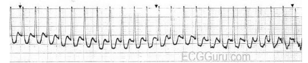
Identify the diagnosis based on the provided ECG image.

A patient presents with chest pain and an ECG showing ST-segment elevation. After treatment, the ECG shows resolution of ST-segment changes, but the patient continues to have chest pain. What is the most likely diagnosis?
In ACLS, which drug is recommended for use after unsuccessful defibrillation attempts following ventricular fibrillation?
Which condition is associated with the ECG pattern known as pseudo P pulmonale?
Explanation: ***Aortic dissection*** - The combination of **sudden, severe chest pain radiating to the back** and a **widened mediastinum** on chest X-ray are classic signs of aortic dissection [1]. - This condition involves a tear in the **aortic intima**, allowing blood to flow between the layers of the aortic wall [1]. *Myocardial infarction* - While it causes **severe chest pain**, it typically presents as a **pressure-like** or **squeezing sensation** and is not usually associated with a widened mediastinum. - The pain is usually localized to the **anterior chest** and may radiate to the left arm or jaw, rather than primarily to the back in this fashion [2]. *Pulmonary embolism* - Presents with **chest pain and dyspnea**, but the pain is often **pleuritic** (worse with breathing) and not typically described as radiating to the back. - A **widened mediastinum** is not a typical finding in pulmonary embolism; instead, a V/Q scan or CT pulmonary angiogram would be diagnostic. *Esophageal rupture* - Can cause **severe retrosternal pain** and may rarely lead to a widened mediastinum due to mediastinitis or hematoma, but it's typically associated with **retching or vomiting** (Boerhaave syndrome) and not explicitly radiating to the back as acutely as described. - The pain from esophageal rupture is distinct from the **tearing pain** of an aortic dissection.
Explanation: The combination of **sudden severe abdominal pain**, a **distended abdomen**, **decreased bowel sounds**, elevated **INR (4.5)** due to warfarin use, and a drop in **hemoglobin (9.1)** strongly indicates active **gastrointestinal bleeding**. [1] - **Holding warfarin** and administering **vitamin K** are immediate critical steps to reverse anticoagulation and stop the bleeding. *Acute pancreatitis; NPO, IV fluids, pain control* - While pancreatitis can cause severe abdominal pain, the **INR of 4.5** and decreased hemoglobin point more directly to a **bleeding diathesis**, which is not a primary feature of pancreatitis. - The absence of typical pancreatitis risk factors (e.g., gallstones, alcohol abuse) and laboratory findings (e.g., elevated amylase/lipase) makes this less likely. *Perforated peptic ulcer; IV antibiotics, surgical consult* - A perforated ulcer typically presents with **peritoneal signs** (e.g., rebound tenderness, rigidity) and often **free air on imaging**, which are not explicitly mentioned in this scenario. [2] - While INR 4.5 is a bleeding risk, it's not the primary cause of perforation itself, though it could exacerbate a bleed from an ulcer. *Acute mesenteric ischemia; urgent surgical intervention required* - **Acute mesenteric ischemia** can cause severe abdominal pain and distention, especially in patients with atrial fibrillation due to potential **embolic events**. - However, the markedly elevated **INR of 4.5** and significant **hemoglobin drop** make primary **GI bleeding** a more direct and immediate concern than ischemia, as the bleeding is actively worsened by the anticoagulation.
Explanation: ***Ischemic heart disease*** - This condition is characterized by **reduced blood flow** to the heart, leading to **myocardial damage**, which can result in left ventricular hypertrophy and systolic dysfunction. - It can cause exertional dyspnea due to compromised heart function and may manifest as swelling in the legs due to heart failure [1][2]. *Hypertrophic cardiomyopathy* - Typically presents with **diastolic dysfunction** rather than systolic dysfunction, characterized by an **increased wall thickness** without the systolic failure observed here. - Symptoms include **syncope** during exertion but are less likely to cause leg swelling. *Aortic stenosis* - Often leads to **concentric left ventricular hypertrophy**; however, it primarily results in **outflow obstruction**, causing symptoms like chest pain and exertional dyspnea rather than systolic dysfunction directly. - Leg swelling is less common and typically occurs later in the course of heart failure due to aortic stenosis. *Dilated cardiomyopathy* - Characterized primarily by **systolic dysfunction** [2] but typically includes signs of **ballooning** of the ventricles rather than merely hypertrophy. - It often results from a variety of factors including **viral infections** or **alcohol abuse**, rather than just ischemic causes.
Explanation: ***Antiplatelet agents*** - **Antiplatelet agents**, specifically **aspirin**, are crucial for long-term management in stable ischemic heart disease to prevent **thrombus formation** and reduce the risk of **myocardial infarction** and **stroke** [1]. - They inhibit **platelet aggregation**, a key step in the pathophysiology of atherothrombotic events. *Calcium channel blockers* - While effective for **symptomatic relief** of angina by causing **vasodilation** and reducing cardiac oxygen demand, they do not directly reduce the risk of **myocardial infarction** in stable ischemic heart disease [3]. - Their primary role is to improve **quality of life** by decreasing frequency and severity of angina attacks [2]. *Nitrates* - **Nitrates** are used for both acute and chronic relief of angina symptoms through **venodilation** and **coronary vasodilation**, which reduce **preload** and improve myocardial oxygen supply [1]. - However, they do not have a proven role in reducing the long-term risk of **myocardial infarction** or improving mortality outcomes in stable CAD. *Beta-blockers* - **Beta-blockers** are indicated for long-term management to reduce **angina symptoms** and improve outcomes after **myocardial infarction** by decreasing heart rate, blood pressure, and myocardial contractility, thereby reducing **myocardial oxygen demand** [3]. - While they reduce MI recurrence post-MI, their primary role in *stable* ischemic heart disease (without prior MI) is for symptom control rather than primary prevention of initial MI beyond their hemodynamic effects.
Explanation: ***HOCM*** - Standing decreases **venous return** and **left ventricular volume**, which reduces the size of the LV outflow tract and thus exacerbates the obstruction in **hypertrophic obstructive cardiomyopathy (HOCM)**, making the murmur louder [1]. - This maneuver is a key diagnostic feature as reduced preload intensifies the dynamic obstruction. *MR* - **Mitral regurgitation (MR)** is typically a volume overload lesion, and standing (which reduces preload) generally causes the murmur to **decrease** in intensity due to less blood volume ejected back into the atrium [3]. - The murmur of MR is usually a holosystolic murmur radiating to the axilla [3]. *MS* - **Mitral stenosis (MS)** is a fixed obstruction to left ventricular filling. Changes in preload (like standing) do not significantly alter the gradient across the mitral valve or the intensity of the murmur [2]. - Its characteristic murmur is a **mid-diastolic rumble** with an opening snap [1]. *VSD* - A **ventricular septal defect (VSD)** murmur is caused by blood flowing from the high-pressure left ventricle to the low-pressure right ventricle. Standing, by reducing systemic vascular resistance, would typically cause the murmur to **decrease** in intensity as less blood shunts left-to-right. - The murmur is usually a **holosystolic murmur** best heard at the lower left sternal border.
Explanation: ***Patent ductus arteriosus*** - **Patent ductus arteriosus (PDA)** is the most characteristic cardiovascular defect associated with congenital rubella syndrome [1]. - The rubella virus can interfere with the normal closure of the **ductus arteriosus** during fetal development [1]. *Coarctation of the aorta* - **Coarctation of the aorta** is a narrowing of the aorta and is not typically linked to congenital rubella syndrome [1]. - It is often associated with syndromes like **Turner syndrome** or bicuspid aortic valve, which are not mentioned here [1]. *Ventricular septal defect* - **Ventricular septal defects (VSDs)** are common congenital heart defects, but they are not specifically characteristic of congenital rubella [1]. - VSDs are more frequently associated with conditions like **Down syndrome** or can occur in isolation [1]. *Pulmonary artery stenosis* - While **pulmonary artery stenosis** can occur as a congenital heart defect, it is less commonly the primary cardiovascular manifestation of congenital rubella syndrome. - **Peripheral pulmonary artery stenosis** can be seen in rubella, but PDA is the most characteristic overarching defect [1].
Explanation: ***PSVT*** - The ECG shows a **narrow complex tachycardia** with a regular rhythm and a high heart rate, characteristic of **paroxysmal supraventricular tachycardia (PSVT)**. - P waves are often **buried within the QRS complex** or T waves, or may be retrograde, which can be seen as small deflections or changes in the baseline in some leads. *VT* - **Ventricular tachycardia** is characterized by a **wide QRS complex** (>0.12 seconds), which is not observed in this ECG. - While VT can be regular, the primary distinguishing feature is the QRS duration. *AT* - **Atrial tachycardia (AT)** is another form of supraventricular tachycardia, but it typically shows **distinct P waves** with an abnormal morphology, often separate from the T wave, which are not clearly visible or consistently distinct in this tracing. - While it can present with narrow complex tachycardia, the mechanism differs from re-entrant PSVT. *Ventricular fibrillation* - **Ventricular fibrillation** is characterized by **chaotic, irregular electrical activity** with no distinguishable P waves, QRS complexes, or T waves, representing disorganized ventricular depolarization. - The ECG in the image shows a consistent, regular rhythm with identifiable, albeit narrow, QRS complexes.
Explanation: ***Takotsubo cardiomyopathy*** - Characterized by **transient ventricular dysfunction** (often apical ballooning) mimicking a heart attack, usually triggered by severe emotional or physical stress. - **Resolution of ST-segment elevation** but persistent chest pain after initial presentation, especially if coronary angiography is normal, strongly suggests this diagnosis. *Acute myocardial infarction* - While initially presenting with **ST-segment elevation** and chest pain, resolution of ST-segment changes without corresponding clinical improvement or complete resolution of symptoms would be unusual without reperfusion, and persistent pain might indicate ongoing ischemia or complications [1], [2]. - The key difference here is the **spontaneous resolution of ST elevation** while pain persists, and the absence of clear coronary artery occlusion in Takotsubo. *Myocarditis* - **Myocarditis** can cause chest pain and ECG abnormalities, including ST-segment elevation, but the specific pattern of resolution of ST changes with persistent pain and the typical trigger of emotional stress are less characteristic. - Diagnosis often involves **cardiac MRI** showing inflammation and is not characterized by the quick resolution of acute ECG changes seen here. *Pericarditis* - Pericarditis typically presents with **pleuritic chest pain** that is relieved by leaning forward, and diffused ST-segment elevation (often PR depression) followed by T-wave inversions. - The **resolution of ST-segment changes** mentioned, especially in the context of an initial ST elevation that might mimic an MI, does not fit the typical evolutionary ECG changes of pericarditis.
Explanation: ***Amiodarone*** - **Amiodarone** is a potent **antiarrhythmic drug** recommended for persistent ventricular fibrillation (VF) or pulseless ventricular tachycardia (VT) unresponsive to initial defibrillation and epinephrine [1]. - It works by blocking potassium channels, prolonging the **repolarization phase** of the action potential, thereby stabilizing the myocardial cell membrane. *Dopamine* - **Dopamine** is a **vasopressor** used to improve blood pressure and cardiac output in cases of symptomatic **bradycardia** unresponsive to atropine or in **shock** with significant hypotension. - It is not indicated for the treatment of ventricular fibrillation [2]. *Adenosine* - **Adenosine** is primarily used to terminate **supraventricular tachycardias (SVTs)** by transiently blocking the AV node. - It is contraindicated in ventricular fibrillation because it can worsen the arrhythmia. *Atropine* - **Atropine** is an **anticholinergic drug** used to treat **symptomatic bradycardia** by increasing heart rate. - It has no role in the management of ventricular fibrillation.
Explanation: ***Hypercalcemia*** - **Hypercalcemia** can cause a characteristic ECG pattern known as pseudo P pulmonale due to its effects on **myocardial repolarization**. - This condition leads to a **shortened QT interval** which causes the T wave to merge with the P wave, giving the appearance of a tall, peaked P wave. *Hypokalemia* - **Hypokalemia** typically presents with **flattened T waves**, prominent U waves, and ST-segment depression on an ECG [1]. - It can prolong repolarization, which is the opposite effect observed with pseudo P pulmonale [1]. *Hyponatremia* - **Hyponatremia** has a less defined direct effect on ECG patterns compared to other electrolyte imbalances. - Severe hyponatremia may lead to **QRS widening** or **bradycardia**, but not pseudo P pulmonale [1]. *Hypocalcemia* - **Hypocalcemia** characteristically causes **prolongation of the QT interval** on an ECG due to delayed ventricular repolarization [2]. - This is distinct from the shortened QT seen in hypercalcemia that contributes to pseudo P pulmonale.
Coronary Artery Disease and Angina
Practice Questions
Acute Coronary Syndromes
Practice Questions
Heart Failure
Practice Questions
Cardiac Arrhythmias
Practice Questions
Valvular Heart Diseases
Practice Questions
Cardiomyopathies
Practice Questions
Pericardial Diseases
Practice Questions
Congenital Heart Disease in Adults
Practice Questions
Hypertension and Hypertensive Emergencies
Practice Questions
Pulmonary Hypertension
Practice Questions
Non-invasive Cardiac Diagnostics
Practice Questions
Preventive Cardiology
Practice Questions
Get full access to all questions, explanations, and performance tracking.
Start For Free