
Cardiology — MCQs

On this page
Which of the following statements is true regarding the rheumatization of the mitral valve?
Recommended interventions to reduce the incidence of coronary artery disease include the following except which of the following?
Asynchronous cardioversion is given in:
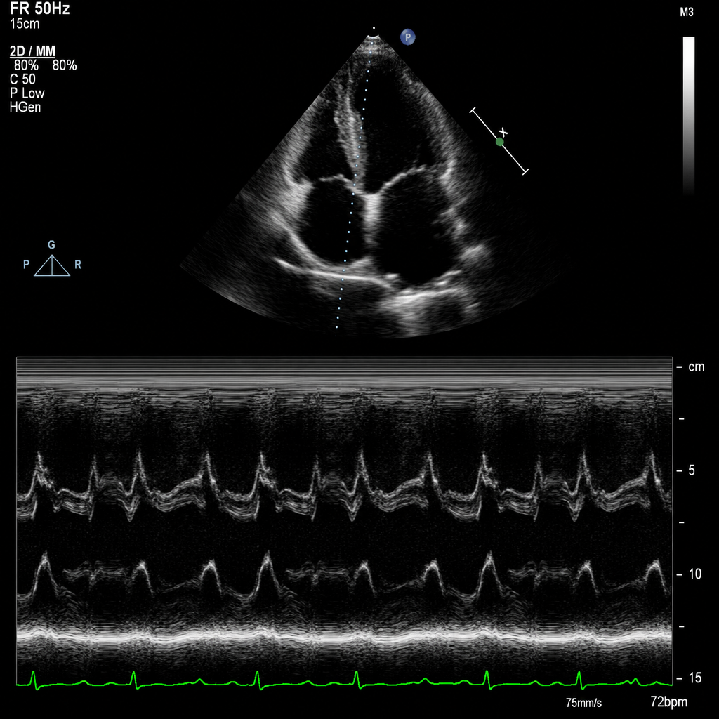
A female patient develops chest pain that is not associated with exercise, and chest auscultation shows multiple non-ejection clicks. The investigation used to diagnose the disease is:
In a patient with congenital prolonged QT syndrome presenting with intermittent torsades de pointes, which of the following is the first-line acute treatment?
Radiofrequency ablation is commonly performed for which of the following conditions?
Which of the following is the primary feature of Eisenmenger syndrome?
ECG is poor in detecting ischemia in areas supplied by which of the following vessels?
Which one of the following is a major criterion in Jones' criteria for rheumatic fever?
A 38-year-old daily laborer has a heart rate of 44 on routine examination. What is the most appropriate management?
Practice by Chapter
Coronary Artery Disease and Angina
Practice Questions
Acute Coronary Syndromes
Practice Questions
Heart Failure
Practice Questions
Cardiac Arrhythmias
Practice Questions
Valvular Heart Diseases
Practice Questions
Cardiomyopathies
Practice Questions
Pericardial Diseases
Practice Questions
Congenital Heart Disease in Adults
Practice Questions
Hypertension and Hypertensive Emergencies
Practice Questions
Pulmonary Hypertension
Practice Questions
Non-invasive Cardiac Diagnostics
Practice Questions
Preventive Cardiology
Practice Questions
Want unlimited practice?
Get full access to all questions, explanations, and performance tracking.
Scan to download app