
Cardiology — MCQs

On this page
Which of the following murmurs increase with a Valsalva maneuver?
ECG of a patient shows more than three consecutive premature ventricular contractions (PVC) with a heart rate of less than 100 beats per minute. What is the diagnosis?
Classification of aortic dissection depends on.
Austin Flint murmur is associated with which condition?
A 54-year-old man presents after a syncopal episode with no recollection of the event, and bystanders report that he regained consciousness approximately 45 seconds after falling. He has a history of bipolar disorder managed with quetiapine, and recently experienced prostatitis treated with ciprofloxacin. His other medications include lisinopril and hydrochlorothiazide for hypertension, and cyclobenzaprine and a hydrocodone/acetaminophen combination pill for low back pain. On examination, the patient is alert and oriented, with a nonfocal neurological examination and an unremarkable cardiac examination. Electrocardiogram shows nonspecific ST and T wave changes and a prolonged QT interval (QTc of 540 milliseconds). What is the best initial management approach?
A patient with new-onset syncope has a blood pressure of 110/95 mmHg and a harsh systolic ejection murmur at the base, radiating to both carotids. What finding may be revealed upon auscultation of the second heart sound at the base?
A 60-year-old woman with a history of diabetes mellitus has had left-sided chest pain radiating to the arm for the past 5 hours. Serial measurements of serum creatine kinase-MB levels show an elevated level 24 hours after the onset of pain. Partial thromboplastin time (PTT) and prothrombin time (PT) are normal. Coronary angiography shows occlusion of the left anterior descending artery. Which of the following mechanisms is the most likely cause of thrombosis in this patient?
A 36-year-old patient presents to the emergency department with shock. His ECG shows ST elevation in leads II and III and aVF. What is the most likely cause of shock?
Which of the following ECG findings is most characteristic of acute myocardial infarction?
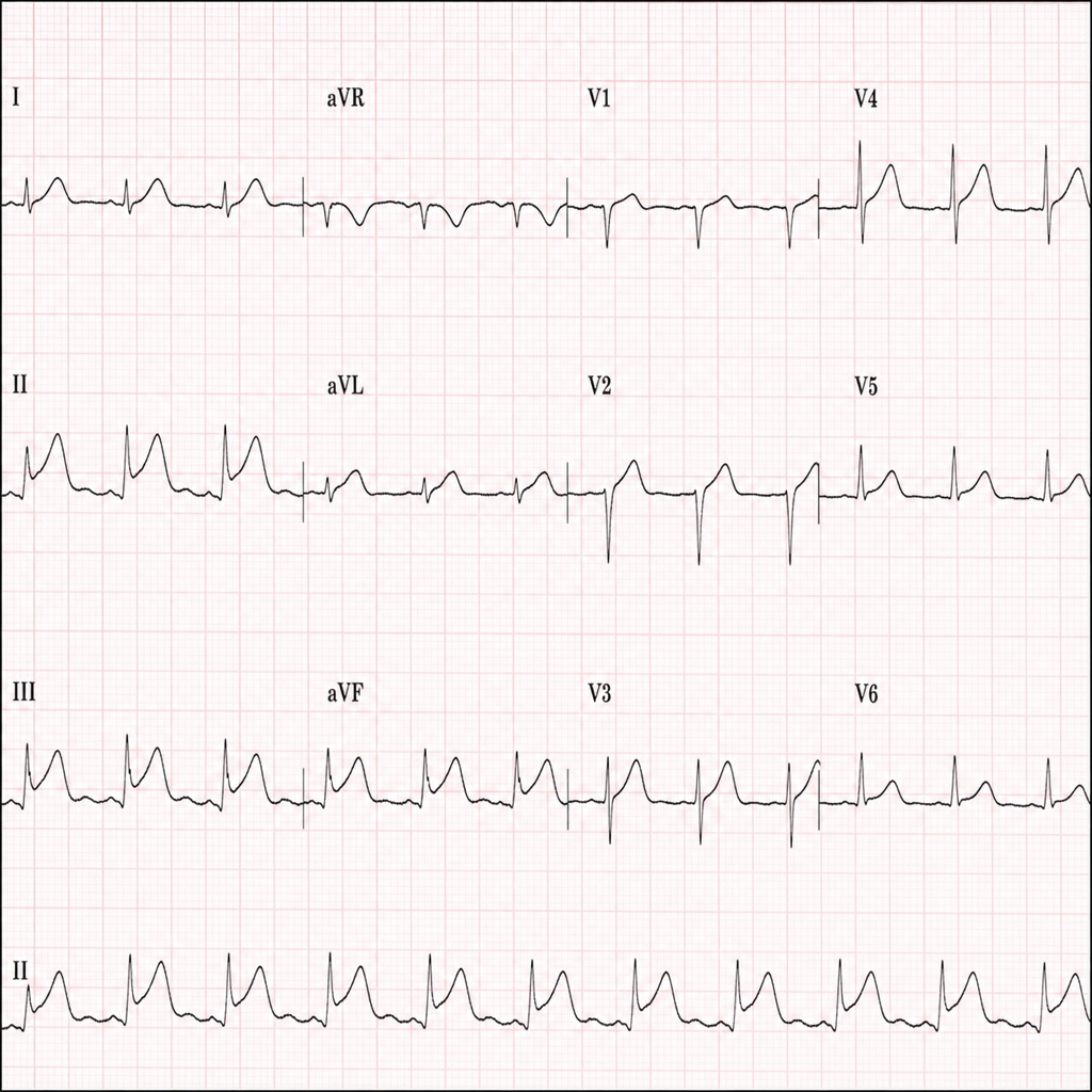
A 68-year-old man presents with a syndrome of alcohol withdrawal. His ECG shows a regular narrow complex tachycardia at 150 bpm with characteristic sawtooth waves. What is the most likely diagnosis?
Practice by Chapter
Coronary Artery Disease and Angina
Practice Questions
Acute Coronary Syndromes
Practice Questions
Heart Failure
Practice Questions
Cardiac Arrhythmias
Practice Questions
Valvular Heart Diseases
Practice Questions
Cardiomyopathies
Practice Questions
Pericardial Diseases
Practice Questions
Congenital Heart Disease in Adults
Practice Questions
Hypertension and Hypertensive Emergencies
Practice Questions
Pulmonary Hypertension
Practice Questions
Non-invasive Cardiac Diagnostics
Practice Questions
Preventive Cardiology
Practice Questions
Want unlimited practice?
Get full access to all questions, explanations, and performance tracking.
Scan to download app