
Cardiology — MCQs

On this page
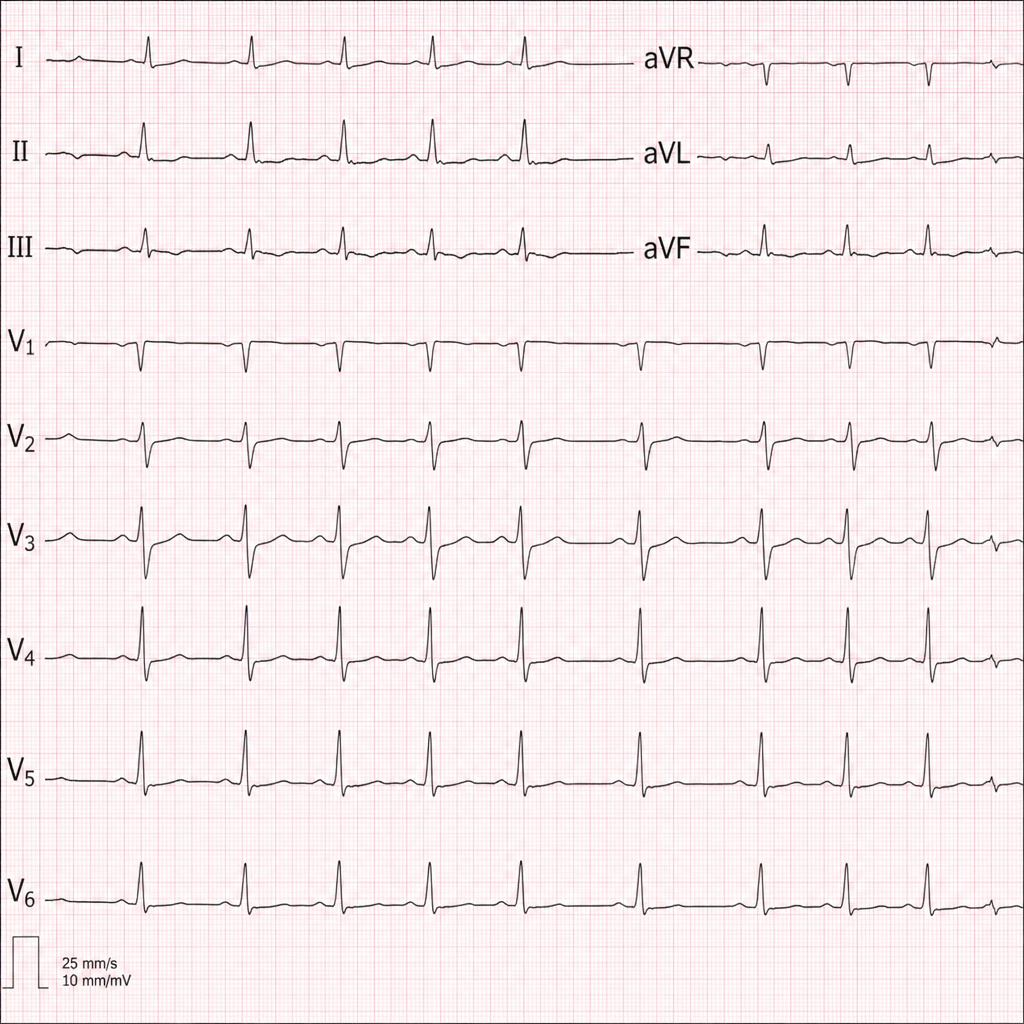
Refer to the provided ECG image. It demonstrates which of the following?
In a patient with heart disease, which condition is most commonly associated with left atrial enlargement?
A 40-year-old male patient presents to the Emergency department with central chest pain for 2 hours. The ECG shows ST segment depression and cardiac troponins are elevated. The patient has a positive history of previous PCI 3 months back. He is administered Aspirin, Clopidogrel, Nitrates, and LMWH in the Emergency Department and shifted to the coronary care unit. What is the best recommended course of further action?
Under which condition are steroids administered in rheumatic fever?
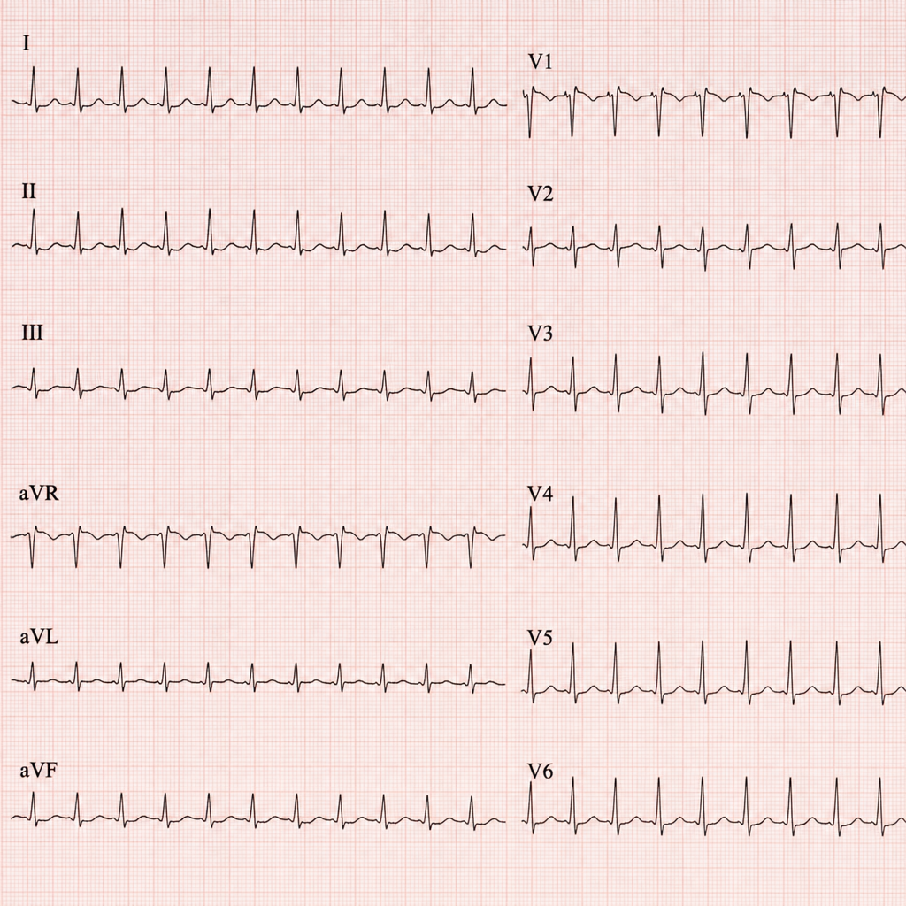
Diagnose the underlying medical disorder based on the ECG changes.
What is the most likely cause of fluid overload in a patient presenting with shortness of breath?
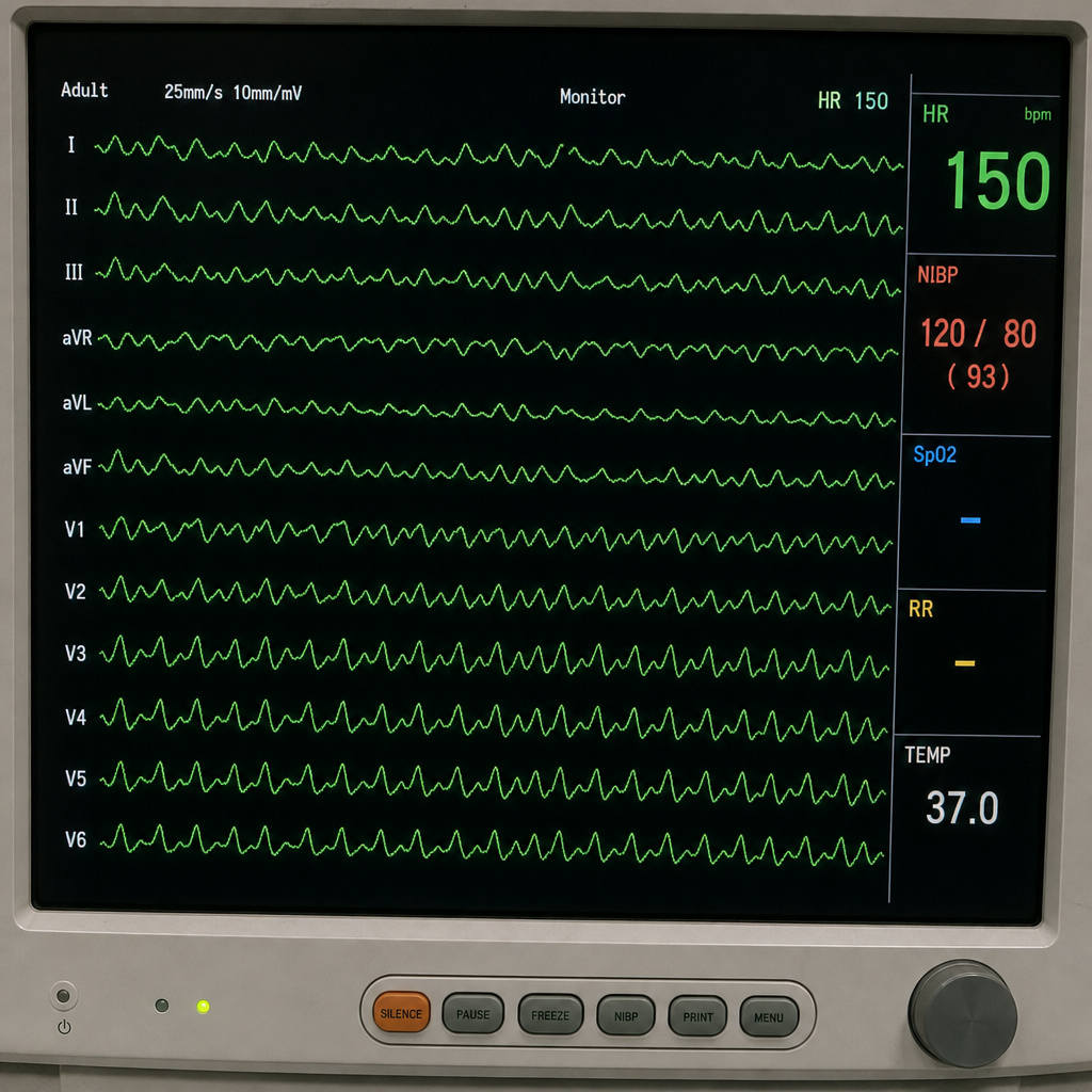
This patient came to the casualty with palpitations. His ECG has been shown below. What is your diagnosis?
Torsades de pointes is seen in all except
Which of the following is associated with WPW syndrome?
Graham Steell murmur is associated with which of the following conditions?
Practice by Chapter
Coronary Artery Disease and Angina
Practice Questions
Acute Coronary Syndromes
Practice Questions
Heart Failure
Practice Questions
Cardiac Arrhythmias
Practice Questions
Valvular Heart Diseases
Practice Questions
Cardiomyopathies
Practice Questions
Pericardial Diseases
Practice Questions
Congenital Heart Disease in Adults
Practice Questions
Hypertension and Hypertensive Emergencies
Practice Questions
Pulmonary Hypertension
Practice Questions
Non-invasive Cardiac Diagnostics
Practice Questions
Preventive Cardiology
Practice Questions
Want unlimited practice?
Get full access to all questions, explanations, and performance tracking.
Scan to download app