
Cardiology — MCQs

On this page
What are the potential causes of cardiogenic shock excluding myocardial infarction?
In which of the following conditions is the implantation of an Automatic Implantable Cardioverter Defibrillator (AICD) indicated?
Roth spots are associated with which of the following conditions?
Which drug is used as an adjunct to epinephrine in refractory ventricular fibrillation/ventricular tachycardia during cardiac arrest?
What does the CEAP score classify?
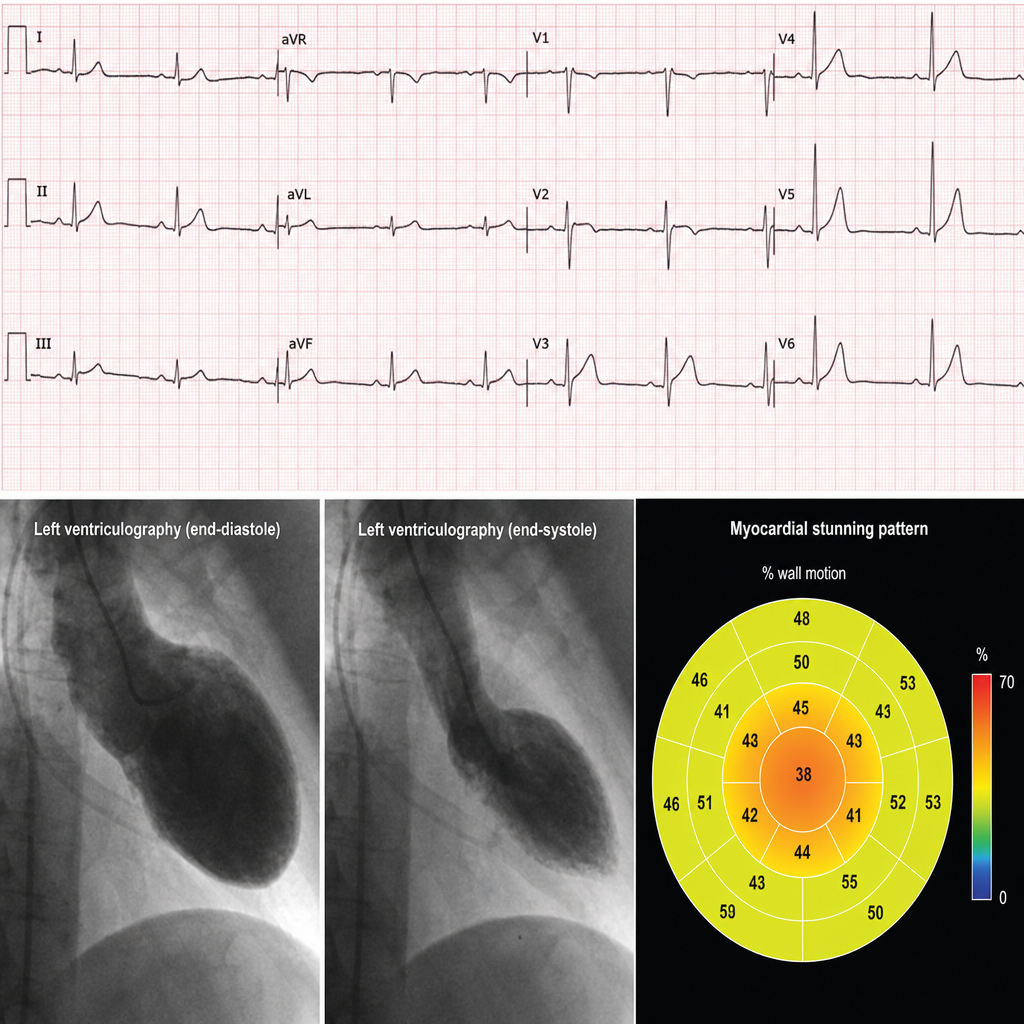
In a patient presenting with an ECG that shows ST-segment elevation but with a myocardial stunning pattern that does not correspond to a single coronary artery territory, what is the most likely diagnosis?
Murmur heard in aortic stenosis
The severity of mitral stenosis can be judged by-
Which of the following is not recommended for patients with coronary artery disease?
In the context of chest pain evaluation, which is the best way to differentiate between stable angina and NSTEMI?
Practice by Chapter
Coronary Artery Disease and Angina
Practice Questions
Acute Coronary Syndromes
Practice Questions
Heart Failure
Practice Questions
Cardiac Arrhythmias
Practice Questions
Valvular Heart Diseases
Practice Questions
Cardiomyopathies
Practice Questions
Pericardial Diseases
Practice Questions
Congenital Heart Disease in Adults
Practice Questions
Hypertension and Hypertensive Emergencies
Practice Questions
Pulmonary Hypertension
Practice Questions
Non-invasive Cardiac Diagnostics
Practice Questions
Preventive Cardiology
Practice Questions
Want unlimited practice?
Get full access to all questions, explanations, and performance tracking.
Scan to download app