
Cardiology — MCQs

On this page
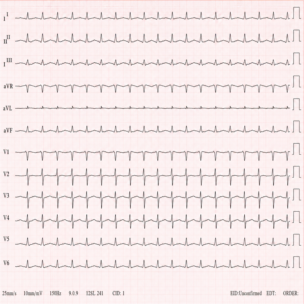
A 32-year-old patient presents to the emergency department with palpitations and dizziness. An ECG is obtained. Identify the cardiac rhythm shown in the image.
Identify the cardiac condition represented in the image.
Which type of cardiomyopathy is associated with alcohol abuse?
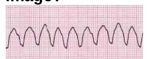
What is the most appropriate immediate management for a hemodynamically unstable patient with supraventricular tachycardia (SVT)?
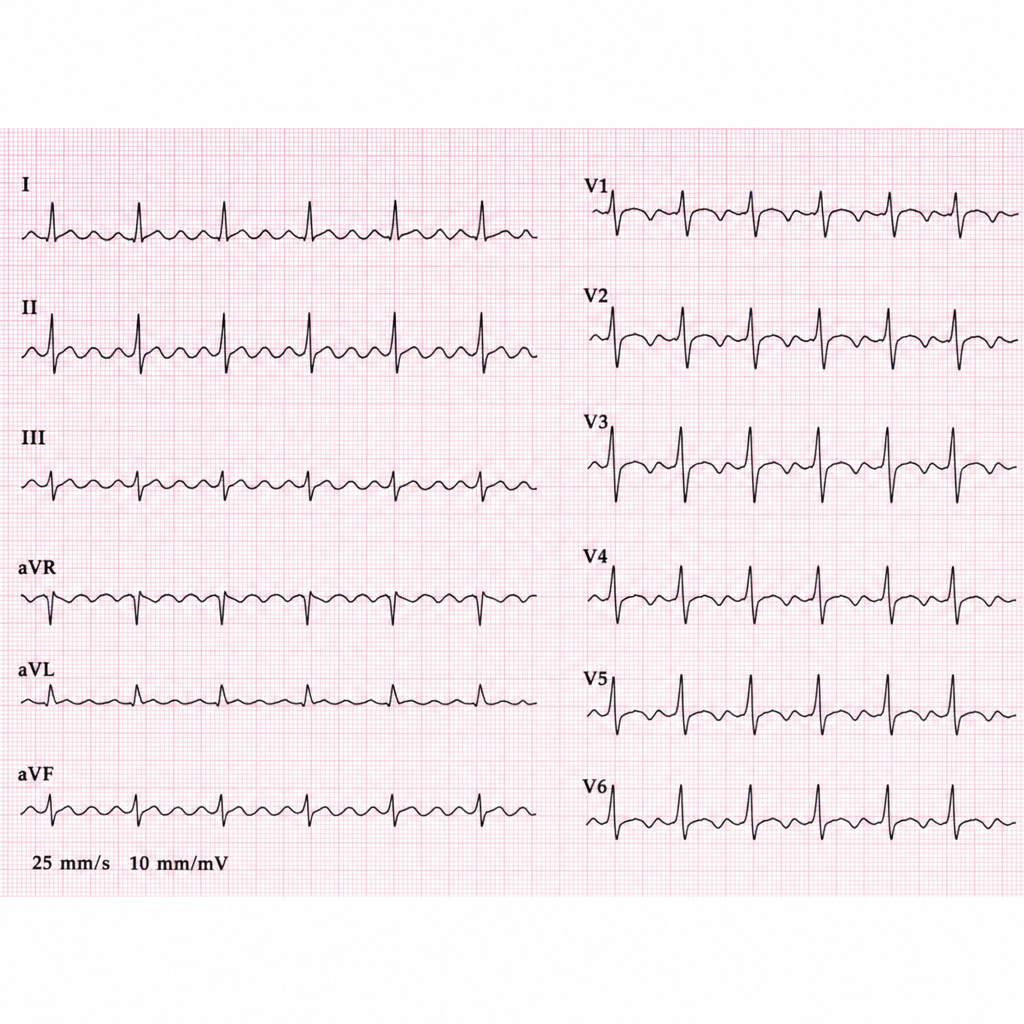
Identify the condition in the provided ECG image.
Osborn J waves are seen in which of the following conditions?
Which of the following statements is true about the Bundle of Kent?
A patient presents to you with an irregularly irregular pulse of 120/minutes and a pulse deficit of 20. Which of the following would be the jugular venous pressure (JVP) finding?
What electrolyte imbalance is most commonly associated with prominent U waves on ECG?
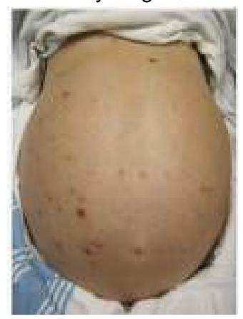
A 55-year-old hypertensive patient presents with severe chest pain radiating to the back. What does a CT scan of the thorax typically reveal?
Practice by Chapter
Coronary Artery Disease and Angina
Practice Questions
Acute Coronary Syndromes
Practice Questions
Heart Failure
Practice Questions
Cardiac Arrhythmias
Practice Questions
Valvular Heart Diseases
Practice Questions
Cardiomyopathies
Practice Questions
Pericardial Diseases
Practice Questions
Congenital Heart Disease in Adults
Practice Questions
Hypertension and Hypertensive Emergencies
Practice Questions
Pulmonary Hypertension
Practice Questions
Non-invasive Cardiac Diagnostics
Practice Questions
Preventive Cardiology
Practice Questions
Want unlimited practice?
Get full access to all questions, explanations, and performance tracking.
Scan to download app