
Cardiology — MCQs

On this page
Which of the following is least likely to be associated with broad complex tachycardia due to ventricular tachycardia?
In Left Ventricular Hypertrophy (LVH), what is the minimum value of SV1 + RV6 in mm that suggests the presence of LVH?
The normal range for Ankle Brachial Pressure Index (ABI) is:
Which of the following is not a high-pitched heart sound?
Which of the following is a complication of Takayasu's arteritis?
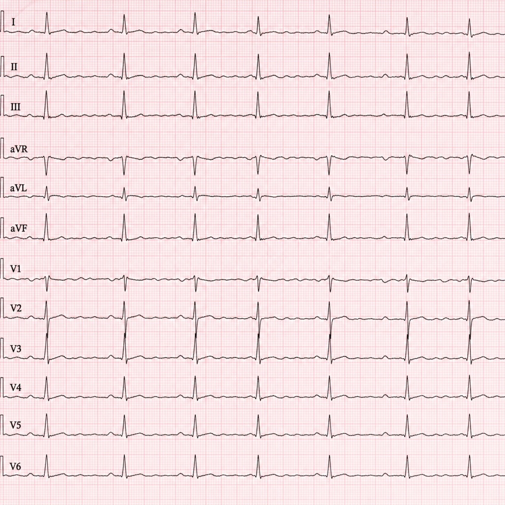
A patient in regular rhythm presents with absent P waves on ECG. Which of the following is the most likely diagnosis based on the ECG findings?
LBBB is seen with all except
Erb's Point in cardiology refers to:
Which of the following is the MOST common complication of untreated hypertension?
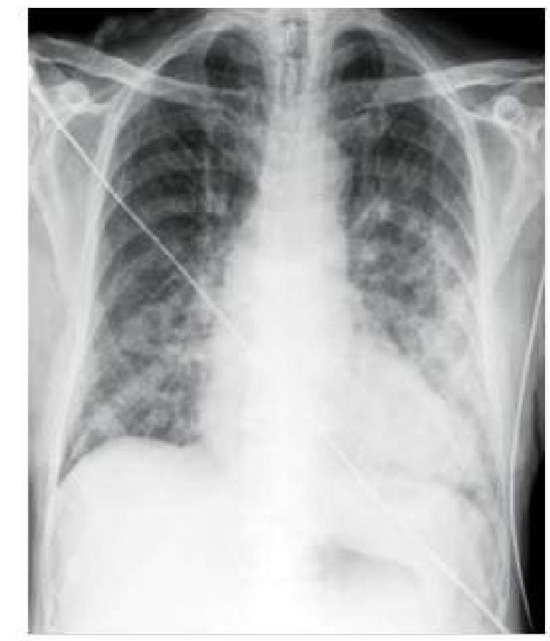
A hypertensive patient who is non-compliant with medication presents to you with sudden onset breathlessness. A chest x-ray was done, which is shown below. How will you manage this patient?
Practice by Chapter
Coronary Artery Disease and Angina
Practice Questions
Acute Coronary Syndromes
Practice Questions
Heart Failure
Practice Questions
Cardiac Arrhythmias
Practice Questions
Valvular Heart Diseases
Practice Questions
Cardiomyopathies
Practice Questions
Pericardial Diseases
Practice Questions
Congenital Heart Disease in Adults
Practice Questions
Hypertension and Hypertensive Emergencies
Practice Questions
Pulmonary Hypertension
Practice Questions
Non-invasive Cardiac Diagnostics
Practice Questions
Preventive Cardiology
Practice Questions
Want unlimited practice?
Get full access to all questions, explanations, and performance tracking.
Scan to download app