
Cardiology — MCQs

On this page
A 45-year-old man presents with exertional dyspnea and swelling of his legs. An echocardiogram shows left ventricular hypertrophy and systolic dysfunction. What is the most likely underlying cause?
Which medication is indicated for the long-term management of patients with stable ischemic heart disease to reduce the risk of myocardial infarction?
Which murmur increases on standing?
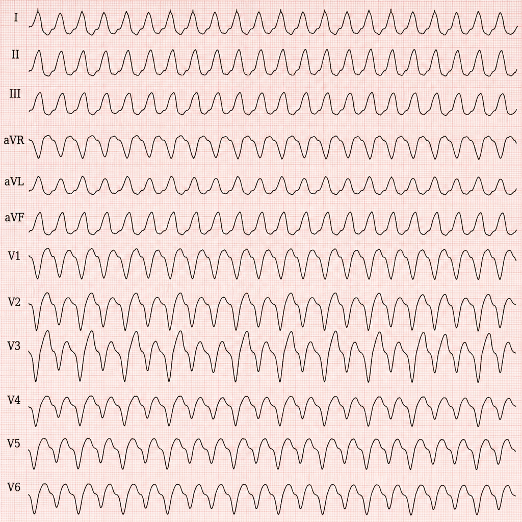
Identify the cardiac condition based on the ECG findings of a wide QRS complex tachycardia.
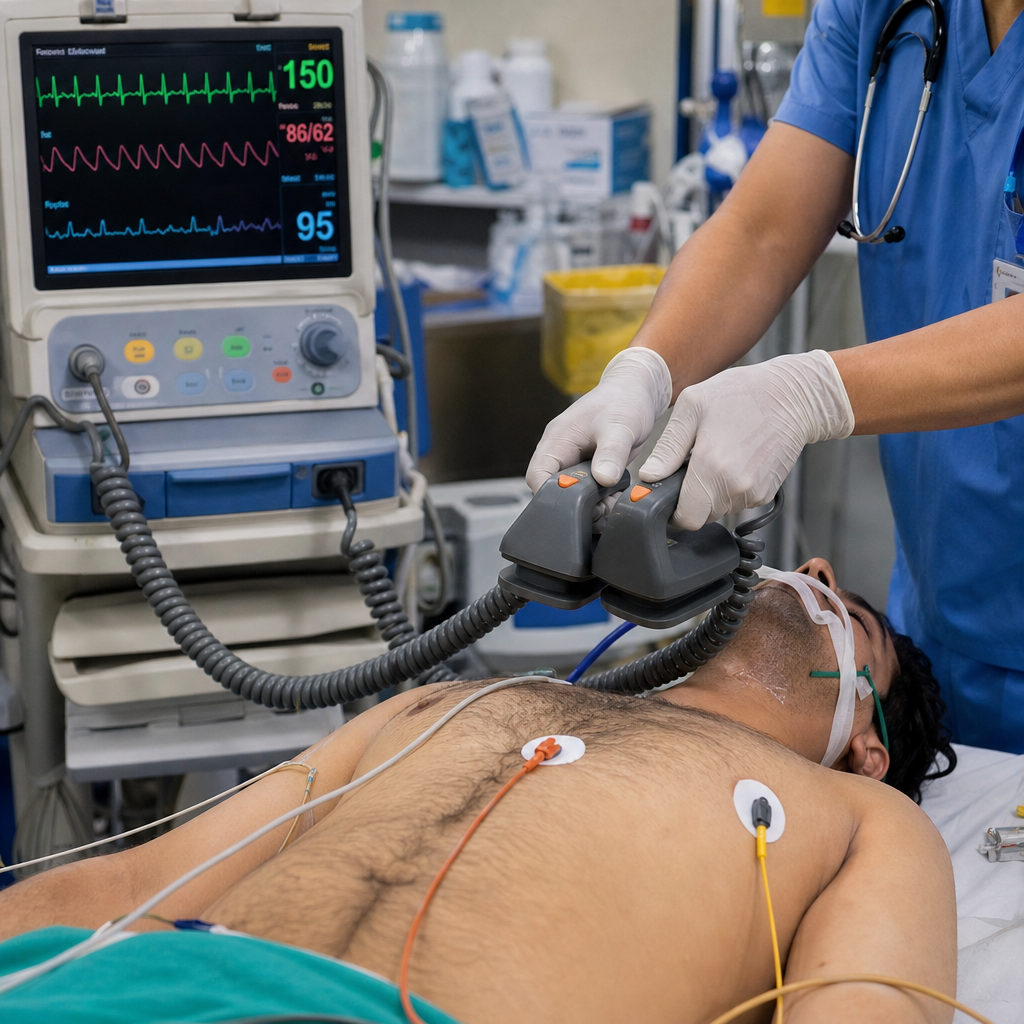
What is the immediate treatment for a hemodynamically unstable patient with supraventricular tachycardia (SVT)?
Identify the diagnosis based on the provided ECG image.
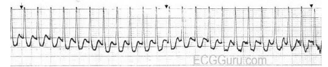
Identify the condition in the ECG based on the provided image.
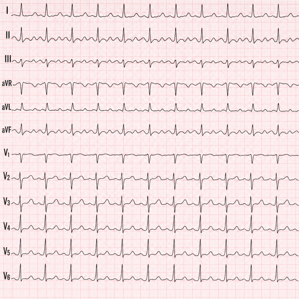
Which condition is associated with the ECG pattern known as pseudo P pulmonale?
What is a potential cause of cardiogenic shock other than myocardial infarction (MI)?
At what time frame does peripartum cardiomyopathy typically occur?
Practice by Chapter
Coronary Artery Disease and Angina
Practice Questions
Acute Coronary Syndromes
Practice Questions
Heart Failure
Practice Questions
Cardiac Arrhythmias
Practice Questions
Valvular Heart Diseases
Practice Questions
Cardiomyopathies
Practice Questions
Pericardial Diseases
Practice Questions
Congenital Heart Disease in Adults
Practice Questions
Hypertension and Hypertensive Emergencies
Practice Questions
Pulmonary Hypertension
Practice Questions
Non-invasive Cardiac Diagnostics
Practice Questions
Preventive Cardiology
Practice Questions
Want unlimited practice?
Get full access to all questions, explanations, and performance tracking.
Scan to download app