
Diseases of the Nose and Paranasal Sinuses — MCQs

On this page
A 72-year-old man presents to the emergency department complaining of frequent nose-bleeds. What is the most likely site of acute epistaxis?
A patient presented to ENT OPD with complaints of headache and nasal stuffiness. On CT scan, heterogeneous opacification involving multiple sinuses along with bone erosion was noticed. What would be the most likely diagnosis?
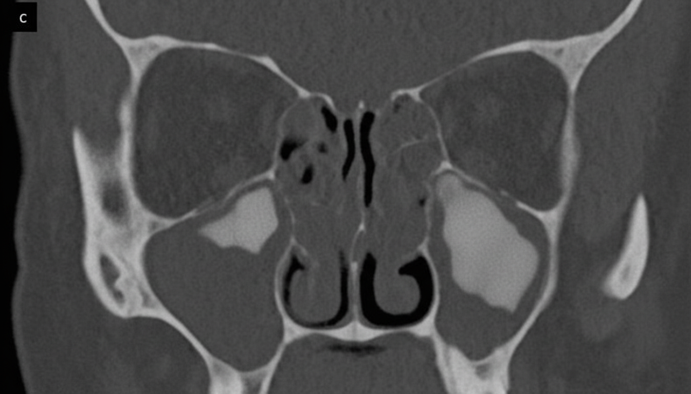
A 16-year-old girl presented with a history of nasal obstruction for the last 2 months. A CT scan was performed, revealing the following findings. What is the most likely diagnosis?
What is the characteristic feature of an ethmoidal polyp?
What is the presenting symptom of nasal myiasis?
Epistaxis in an elderly person is most commonly due to:
A diabetic male presents with facial pain and blackish discoloration in the nose. The CT image shows bone erosion and sinus involvement. What is the most likely diagnosis?
Which of the following is shown in the image below (circled)?
A patient presented to the OPD with complaints of greenish black matter in the nose with foul-smelling discharge. What is the diagnosis?
A 45-year-old male was admitted with respiratory distress. CT showed a nasal polyp with fluid collection in the sinus. Drainage of which of the following is obstructed?
Practice by Chapter
Rhinitis
Practice Questions
Acute Rhinosinusitis
Practice Questions
Chronic Rhinosinusitis
Practice Questions
Nasal Polyposis
Practice Questions
Allergic Fungal Sinusitis
Practice Questions
Deviated Nasal Septum
Practice Questions
Epistaxis
Practice Questions
Nasal Trauma
Practice Questions
Choanal Atresia
Practice Questions
CSF Rhinorrhea
Practice Questions
Tumors of the Nose and Paranasal Sinuses
Practice Questions
Complications of Sinusitis
Practice Questions
Want unlimited practice?
Get full access to all questions, explanations, and performance tracking.
Scan to download app