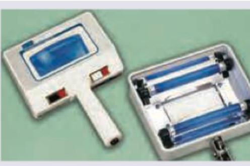
Which of the following are uses of Wood's light examination?
Tzanck preparation is used for the diagnosis of which of the following skin conditions, EXCEPT?
Complete circumferential and peripheral deep margin assessment is known as:
What is the best treatment for achieving cosmetic results in large port-wine hemangiomas?
Mohs surgery is indicated for which of the following conditions?
What is the wavelength of the carbon dioxide laser?
Microdermabrasion is done with:
The first line treatment for this condition is:

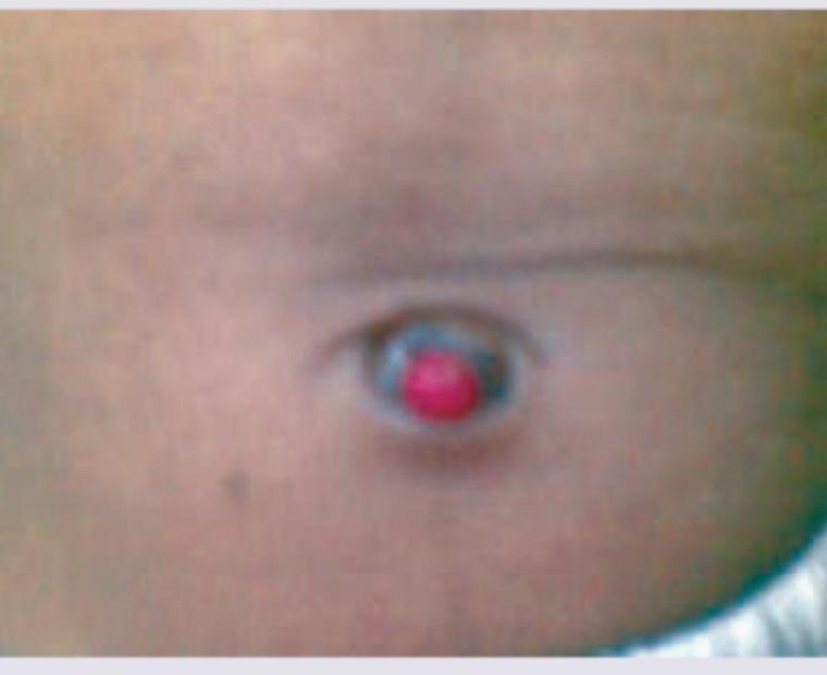
The following image shows:

Best treatment for the lesion shown in the figure?

Explanation: **Explanation:** Wood’s light examination is a diagnostic tool that uses long-wave ultraviolet light (365 nm) to detect specific fluorescence in skin and appendages, aiding in the diagnosis of various dermatological conditions. * **Urine examination in Porphyria (Option A):** In Porphyria Cutanea Tarda (PCT), the urine contains high levels of uroporphyrins. When exposed to Wood’s light, the urine exhibits a characteristic **coral-red or pink fluorescence**. * **Examination of hair in Tinea capitis (Option B):** Wood’s light is used to screen for fungal infections. Specifically, **Microsporum** species (like *M. audouinii* and *M. canis*) produce a **bright blue-green** fluorescence due to the presence of pteridine. Note: *Trichophyton schoenleinii* produces a dull greenish fluorescence. * **Sclerema (Option C):** In Sclerema neonatorum, Wood’s light is used to detect the presence of subcutaneous fat necrosis or specific metabolic changes, though this is a less common but academically recognized application in older literature. **Clinical Pearls for NEET-PG:** * **Erythrasma:** Shows a pathognomonic **coral-red** fluorescence (due to Coproporphyrin III from *Corynebacterium minutissimum*). * **Pseudomonas:** Shows **yellow-green** fluorescence (due to pyoverdin). * **Vitiligo:** Shows **milky-white** fluorescence (due to complete loss of melanin, which enhances auto-fluorescence of dermal collagen). * **Pityriasis Versicolor:** Shows **yellowish-orange/copper-orange** fluorescence. Since all listed options represent valid clinical applications of Wood's light, **Option D** is the correct answer.
Explanation: ### Explanation The **Tzanck smear** is a rapid bedside diagnostic test used primarily to identify **acantholytic cells** (Tzanck cells) and multinucleated giant cells. It involves taking a scraping from the base of a fresh vesicle or bulla. **Why Fungal Infections is the Correct Answer:** Tzanck preparation is not used for diagnosing fungal infections. The gold standard bedside test for fungal elements (hyphae, spores, or budding yeast) is a **KOH (Potassium Hydroxide) mount**, which dissolves keratin to visualize the fungi. Tzanck smear lacks the specific stains or chemical properties required to identify fungal morphology effectively. **Analysis of Other Options:** * **Pemphigus (Option A):** This is the classic indication for a Tzanck smear. It reveals **acantholytic cells** (rounded keratinocytes with hyperchromatic nuclei) due to the loss of intercellular adhesion. * **Stevens-Johnson Syndrome (Option B):** In SJS/TEN, a Tzanck smear shows **necrotic keratinocytes** and inflammatory cells, helping to differentiate it from staphylococcal scalded skin syndrome (SSSS). * **Senear-Usher Syndrome (Option C):** Also known as **Pemphigus Erythematosus**, it is a localized variant of Pemphigus Foliaceus. Since it is an immunobullous disease characterized by acantholysis, Tzanck cells will be present. **High-Yield Clinical Pearls for NEET-PG:** 1. **Herpes Simplex/Varicella Zoster:** Tzanck smear shows **Multinucleated Giant Cells** (with Cowdry A inclusion bodies). 2. **Molluscum Contagiosum:** Shows large, oval, eosinophilic cytoplasmic inclusion bodies known as **Henderson-Paterson bodies**. 3. **Donovanosis (Granuloma Inguinale):** Shows **Donovan bodies** (safety-pin appearance) within macrophages. 4. **Staphylococcal Scalded Skin Syndrome (SSSS):** Tzanck smear shows acantholytic cells (similar to Pemphigus), but the cleavage is more superficial (subcorneal).
Explanation: **Explanation:** **Mohs Micrographic Surgery (MMS)** is the gold standard for treating high-risk skin cancers (like Basal Cell Carcinoma and Squamous Cell Carcinoma) because it offers the highest cure rate while preserving maximal healthy tissue. The defining feature of Mohs surgery is the **Complete Circumferential Peripheral and Deep Margin Assessment (CCPDMA)**. Unlike standard wide local excision, where only 1% of the margin is typically examined using "bread-loafing" sections, Mohs surgery involves flattening the tissue specimen so that 100% of the peripheral and deep surgical margins are visualized under the microscope. This ensures that any "roots" or subclinical extensions of the tumor are identified and removed in real-time. **Analysis of Options:** * **Option A (Mohs Surgery):** Correct. It utilizes the CCPDMA technique to ensure total margin control. * **Option B (SHAWN's procedure):** Incorrect. There is no recognized dermatological procedure by this name. It is likely a distractor or a misspelling of "Slow Mohs" (a variation used for melanoma), but it does not represent the standard terminology for margin assessment. * **Options C & D:** Incorrect based on the validity of Option A. **High-Yield Clinical Pearls for NEET-PG:** * **Indications for Mohs:** Tumors in "H-zone" of the face (nose, eyelids, ears), recurrent tumors, ill-defined borders, and aggressive histological subtypes (e.g., morpheaform BCC). * **Tissue Processing:** Uses **frozen sections**, allowing for rapid diagnosis while the patient waits. * **Advantage:** Provides the lowest recurrence rate (approx. 1-2%) and is tissue-sparing, which is critical for cosmetically sensitive areas.
Explanation: **Explanation:** **Port-wine stains (PWS)**, also known as nevus flammeus, are congenital capillary malformations. The gold standard for treatment is **Laser ablation**, specifically using the **Pulsed Dye Laser (PDL)**. The underlying medical concept is **Selective Photothermolysis**. The PDL emits a wavelength (typically 585 or 595 nm) that is specifically absorbed by oxyhemoglobin within the dilated capillaries. This generates heat that destroys the vessel walls while sparing the surrounding dermis and epidermis. This selectivity is crucial for achieving excellent cosmetic results with minimal scarring, especially in large lesions. **Why other options are incorrect:** * **Excision and split-thickness skin graft:** This is highly invasive and often results in significant scarring, texture mismatch, and poor cosmetic outcomes compared to laser therapy. * **Cryosurgery:** This involves non-specific tissue destruction using extreme cold. It lacks selectivity, carries a high risk of permanent pigmentary changes (hypopigmentation), and often leads to hypertrophic scarring. * **Tattooing:** While it can mask the color by injecting skin-toned pigments, it does not treat the underlying vascular pathology. Over time, the pigment may shift or fade unevenly, and it makes subsequent laser treatment impossible. **High-Yield Clinical Pearls for NEET-PG:** * **Drug of Choice (Laser):** Pulsed Dye Laser (PDL) is the first line. * **Wavelength:** 585–595 nm (Yellow light). * **Associated Syndromes:** Always rule out **Sturge-Weber Syndrome** (if the PWS involves the V1/V2 distribution of the trigeminal nerve) and **Klippel-Trenaunay Syndrome** (if involving a limb with hypertrophy). * **Early Intervention:** Treatment is most effective when started in infancy because the skin is thinner and the lesions are smaller.
Explanation: **Explanation:** Mohs Micrographic Surgery (MMS) is a specialized, tissue-sparing surgical technique designed to provide the highest possible cure rate while minimizing the loss of healthy tissue. The core concept of Mohs surgery is the **intraoperative microscopic examination** of 100% of the peripheral and deep surgical margins. * **Basal Cell Carcinoma (BCC):** This is the most common indication for Mohs surgery, particularly for tumors in "high-risk" areas (the H-zone of the face: nose, eyelids, lips, ears) or aggressive histological subtypes (morpheaform or micronodular). * **Squamous Cell Carcinoma (SCC):** Mohs is indicated for SCCs that are recurrent, large (>2cm), poorly differentiated, or located in cosmetically sensitive areas where tissue preservation is vital. * **Melanoma:** While traditionally treated with wide local excision, **Mohs surgery (specifically using MART-1 immunostaining)** is increasingly used for Melanoma in situ (Lentigo Maligna) to ensure clear margins in areas where subclinical extension is common. **Why "All of the above" is correct:** While BCC is the most frequent application, the technique is fundamentally indicated for any skin malignancy where tissue conservation is critical and the tumor borders are difficult to clinically demarcate. **Clinical Pearls for NEET-PG:** * **Gold Standard:** Mohs surgery offers the highest cure rate for primary BCC (~99%) and recurrent BCC (~94%). * **The "H-Zone":** High-risk areas of the face where Mohs is most preferred (Nose, Periorbital, Temple, Ear, Lip). * **Technique:** The tissue is frozen, sectioned horizontally (mapping), and examined by the surgeon themselves, acting as both surgeon and pathologist. * **Other Indications:** Dermatofibrosarcoma Protuberans (DFSP) and Microcystic Adnexal Carcinoma.
Explanation: **Explanation:** The **Carbon Dioxide (CO₂) laser** is a gas laser that operates in the far-infrared spectrum at a wavelength of **10,600 nm**. Its primary chromophore is **water**, which is abundant in biological tissues. Because water highly absorbs this wavelength, the laser energy causes rapid heating and vaporization of intracellular water, leading to precise tissue ablation with minimal collateral thermal damage. **Analysis of Options:** * **10,600 nm (Correct):** The standard wavelength for CO₂ lasers. It is widely used in dermatology for treating benign growths (warts, seborrheic keratosis), skin resurfacing, and "bloodless" surgical incisions. * **1082 nm (Incorrect):** This is not a standard dermatological laser wavelength. * **2940 nm (Incorrect):** This is the wavelength of the **Er:YAG (Erbium-doped Yttrium Aluminum Garnet) laser**. Like the CO₂ laser, its chromophore is water, but it is absorbed 10–15 times more efficiently, allowing for even more superficial and precise ablation. * **1064 nm (Incorrect):** This is the wavelength of the **Nd:YAG laser**. It is used for deep vascular lesions, hair removal in darker skin types (Fitzpatrick IV-VI), and tattoo removal (Q-switched). **High-Yield Clinical Pearls for NEET-PG:** * **Chromophore:** For CO₂ and Er:YAG, the chromophore is **Water**. * **Mode of Action:** CO₂ lasers can be used in "continuous wave" for cutting or "fractionated" mode for skin rejuvenation (Fractional CO₂). * **Safety:** CO₂ laser plumes can contain viable viral particles (e.g., HPV); hence, a smoke evacuator is mandatory during procedures like wart removal. * **Gold Standard:** The CO₂ laser remains the gold standard for traditional laser skin resurfacing.
Explanation: **Explanation:** Microdermabrasion is a non-invasive, superficial mechanical peeling procedure used to exfoliate the stratum corneum. It works on the principle of **kinetic energy**, where abrasive particles are propelled against the skin surface and subsequently removed via vacuum suction. **1. Why Aluminium Oxide is Correct:** **Aluminium oxide (Alumina)** is the gold standard abrasive used in microdermabrasion. It is preferred because it is chemically inert, extremely hard (second only to diamond), and has an irregular crystalline structure that provides superior abrasive properties. It does not cause allergic reactions or systemic toxicity, making it safe for resurfacing. **2. Analysis of Incorrect Options:** * **Aluminium trihydrate crystals:** While sometimes used as a softer alternative for sensitive skin, it is less effective than aluminium oxide and is not the standard material for the procedure. * **Ferrous/Ferric oxide crystals:** These are iron oxides. They are primarily used as pigments (e.g., in calamine or tattoos) or polishing agents in industrial settings, but they are not used in dermatological microdermabrasion due to potential skin staining and lack of appropriate abrasive geometry. **Clinical Pearls for NEET-PG:** * **Indications:** Acne scars (superficial), photoaging, striae distensae, and melasma. * **Depth of Action:** It targets only the **stratum corneum**. * **Alternative Methods:** Besides crystals, "Diamond-tip" microdermabrasion is a popular crystal-free alternative. * **Post-procedure care:** Strict photoprotection is mandatory as the skin's natural barrier is temporarily compromised. * **Contraindications:** Active viral infections (Herpes simplex, warts), active acne (inflammatory), and recent use of Isotretinoin (within 6 months).
Explanation: ***Injection of triamcinolone*** - The image shows a **keloid**, which is an elevated scar that extends beyond the original wound boundaries. Intralesional injections of **corticosteroids** like triamcinolone are considered a first-line treatment for keloids, reducing inflammation and fibroblast proliferation. - This treatment aims to flatten the keloid, relieve symptoms like itching and pain, and prevent recurrence. *Wide excision and grafting* - **Wide excision** of keloids often leads to **recurrence** that is usually larger than the original keloid, making it an unsuitable primary treatment unless combined with other therapies like radiation or intralesional injections. - **Skin grafting** is generally reserved for very large defects after excision and doesn't prevent recurrence of the keloid itself. *Silicon gel sheeting* - **Silicone gel sheeting** or topical silicone is a commonly used non-invasive treatment for scars, particularly **hypertrophic scars** and smaller keloids. - While effective in some cases for preventing and managing scars, it is generally considered a second-line or adjunctive therapy for established keloids, less effective than intralesional corticosteroids for reducing their size. *No intervention* - **Keloids** are **dermatological conditions** that can cause cosmetic disfigurement, itching, pain, and discomfort. - **No intervention** would mean that the keloid would likely persist or even continue to grow, leading to ongoing symptoms and aesthetic concerns.
Explanation: ***Wood's lamp*** - The image clearly displays a device with **ultraviolet (UV) light bulbs** (blue tubes) and a magnifying lens, which is characteristic of a Wood's lamp. - A Wood's lamp is used in dermatology to detect various skin conditions by observing the **fluorescence** of certain substances under UV light. *Infrared lamp* - An infrared lamp produces **heat** and is typically used for therapeutic purposes such as pain relief, not for diagnostic skin examination using UV fluorescence. - Its appearance would involve a bulb that emits visible red light and heat, not the distinct blue UV tubes seen here. *Slit lamp* - A slit lamp is a **biomicroscope** used in ophthalmology to examine the anterior segment and posterior segment of the human eye. - It consists of a light source that can be focused into a narrow slit and a binocular microscope, which looks completely different from the device pictured. *Thermoprobe* - A thermoprobe is a device used to measure **temperature**, often a small probe with a digital readout. - It has no magnifying capabilities or UV light sources, making it distinct from the instrument shown in the image.
Explanation: ***Silver nitrate cauterization*** - The image shows an **umbilical granuloma**, a common benign condition appearing as a soft, reddish-pink moist lesion in the umbilical region after the umbilical cord stump falls off. - **Silver nitrate cauterization** is the gold standard treatment for umbilical granulomas due to its effectiveness in drying and shrinking the granuloma with minimal invasiveness. *Wait and watch for spontaneous regression* - While some mild cases of umbilical granuloma might resolve spontaneously, this lesion appears significant enough to warrant intervention, especially if it's persistent or causing discharge. - **Spontaneous regression** is less likely for larger, more symptomatic granulomas, and waiting can prolong discomfort or risk infection. *Fistulogram and surgical excision* - A **fistulogram and surgical excision** would be indicated if there's suspicion of a **patent omphalomesenteric duct** or a **urachal fistula**, which are much more serious conditions. - The image does not present features suggestive of a fistula (e.g., persistent fecal or urinary discharge), making surgical excision an overly aggressive approach for a simple granuloma. *Laser excision* - **Laser excision** is a viable but usually **second-line treatment** for umbilical granulomas, often reserved for cases that fail silver nitrate treatment or are larger. - It is more invasive and typically more expensive than silver nitrate cauterization, which is effective for most cases.
Skin Biopsy Techniques
Practice Questions
Cryotherapy
Practice Questions
Electrosurgery
Practice Questions
Curettage and Electrodessication
Practice Questions
Excisional Surgery
Practice Questions
Mohs Micrographic Surgery
Practice Questions
Chemical Peels
Practice Questions
Dermabrasion and Microdermabrasion
Practice Questions
Laser Therapy Basics
Practice Questions
Injectable Fillers and Botulinum Toxin
Practice Questions
Photodynamic Therapy
Practice Questions
Wound Care and Dressings
Practice Questions
Get full access to all questions, explanations, and performance tracking.
Start For Free