
Bacterial Skin Infections — MCQs

On this page
Erythematous lesions seen in the axilla with coral red fluorescence under a Wood's lamp indicate which of the following conditions?
Exacerbation of lesions in patients of borderline leprosy is seen in which type of reaction?
Satellite lesions are seen in which type of leprosy?
Which of the following conditions is characterized by 'honey-colored' crusts?
A patient presents with bilateral tender lymphadenopathy and a history of sexual contact. The patient is a truck driver by profession. What is the probable causative agent?
Which of the following is NOT a characteristic of the early eruption of secondary syphilis?
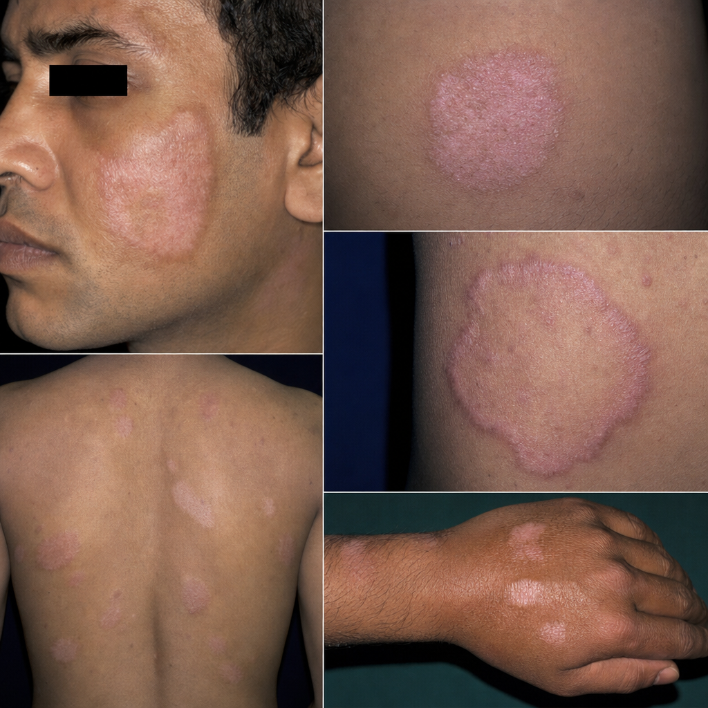
Which type of leprosy is characterized by these lesions?
Wood's lamp examination in dermatology typically reveals a coral red fluorescence in which of the following conditions?
A 20-year-old male presents with multiple painful ulcers over the prepuce and glans, accompanied by suppurative lymphadenopathy, developing two weeks after unprotected sexual intercourse. What is the most probable diagnosis?
What is the recommended treatment for granuloma inguinale?
Practice by Chapter
Impetigo
Practice Questions
Folliculitis, Furuncles, and Carbuncles
Practice Questions
Ecthyma
Practice Questions
Erysipelas and Cellulitis
Practice Questions
Staphylococcal Scalded Skin Syndrome
Practice Questions
Necrotizing Fasciitis
Practice Questions
Cutaneous Tuberculosis
Practice Questions
Leprosy
Practice Questions
Lyme Disease
Practice Questions
Syphilis
Practice Questions
Antibiotic Resistance in Dermatology
Practice Questions
Prophylaxis and Management
Practice Questions
Want unlimited practice?
Get full access to all questions, explanations, and performance tracking.
Scan to download app