
Bacterial Skin Infections — MCQs

On this page
Which of the following is NOT a typical presenting feature of Gonorrhoea?
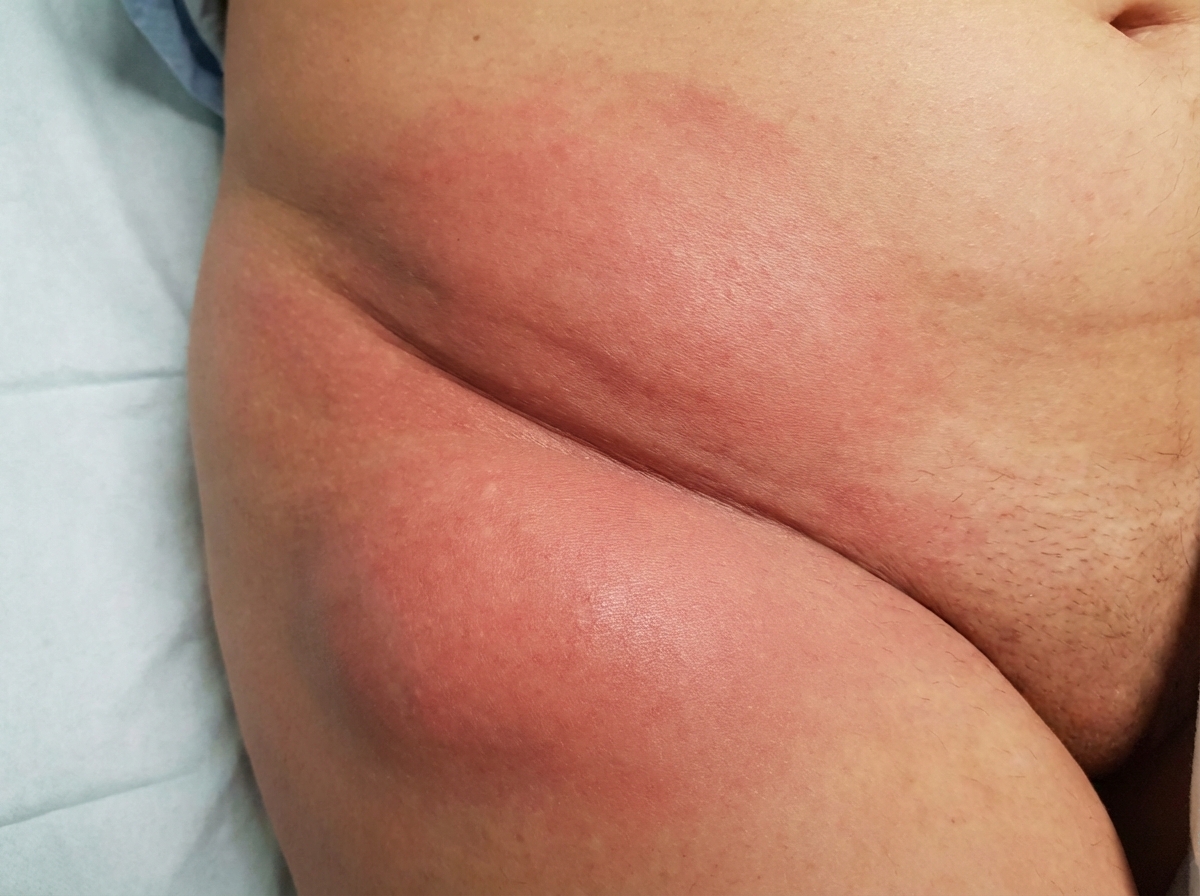
The inguinal region shown in the image is characteristic of which of the following conditions?
A 12-year-old boy presented with a gradually progressive plaque on his buttock for the last 3 years. The plaque was 15 cm in diameter, annular in shape, with crusting and induration at the periphery and scarring at the center. What is the most likely diagnosis?
A 3-year-old child presents with multiple isolated lesions on the face and neck. Physical examination reveals lesions up to 4 cm in diameter with golden crusts, and in other areas, small blisters and weeping are observed. What is the most likely diagnosis?
Gonococcal vaginitis occurs in:
Which of the following is NOT true regarding erysipelas?
Which of the following is a characteristic feature of Borderline leprosy?
Which of the following drugs is not used in the management of lepra reactions?
A young male presented with a hypoesthetic patch on the right forearm. On examination, a thickened peripheral nerve was palpable. Skin biopsy histopathology shows well-formed epithelioid granulomas with Langhans giant cells, dense lymphocytic infiltrate, and perineural infiltration. What is the most likely diagnosis?
A young female presented to the hospital OPD with suspected chlamydial infection. She was prescribed oral doxycycline for 2 weeks. After 3 weeks, she came to the hospital again with a mucopurulent cervicitis. On questioning, she admitted that she took the drug only for 3 days. What should be done next?
Practice by Chapter
Impetigo
Practice Questions
Folliculitis, Furuncles, and Carbuncles
Practice Questions
Ecthyma
Practice Questions
Erysipelas and Cellulitis
Practice Questions
Staphylococcal Scalded Skin Syndrome
Practice Questions
Necrotizing Fasciitis
Practice Questions
Cutaneous Tuberculosis
Practice Questions
Leprosy
Practice Questions
Lyme Disease
Practice Questions
Syphilis
Practice Questions
Antibiotic Resistance in Dermatology
Practice Questions
Prophylaxis and Management
Practice Questions
Want unlimited practice?
Get full access to all questions, explanations, and performance tracking.
Scan to download app