
Bacterial Skin Infections — MCQs

On this page
What is the most likely diagnosis for a patient presenting with a painful ulcer on the glans penis?
A 23-year-old presented with a painless penile ulcer and painless lymphadenopathy. What is the diagnosis?
Which of the following is NOT involved in leprosy?
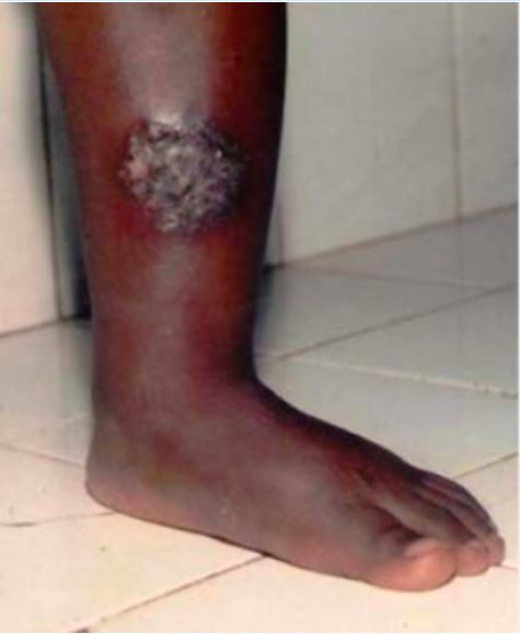
A farmer presents with a lesion on the leg. Which of the following is the most likely diagnosis?
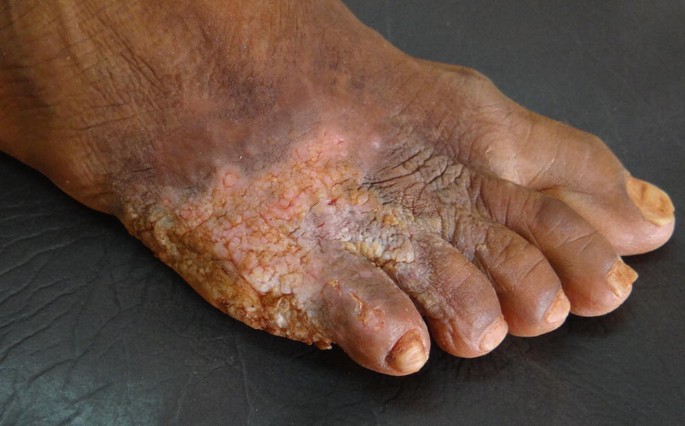
Apple jelly nodules on diascopy are characteristic of which condition?
What is the recommended duration for the treatment of multibacillary leprosy?
A farmer presents with a lesion on the leg. Which of the following is the most likely diagnosis?
In a diagnosed lepromatous leprosy patient, treatment was started. After intake of the drug, skin lesions and fever develop within a few days. Which type of hypersensitivity reaction leads to this manifestation?
What is the best method of treatment for ulnar nerve abscess in case of leprosy?
Which of the following tests is NOT used for the diagnosis of leprosy?
Practice by Chapter
Impetigo
Practice Questions
Folliculitis, Furuncles, and Carbuncles
Practice Questions
Ecthyma
Practice Questions
Erysipelas and Cellulitis
Practice Questions
Staphylococcal Scalded Skin Syndrome
Practice Questions
Necrotizing Fasciitis
Practice Questions
Cutaneous Tuberculosis
Practice Questions
Leprosy
Practice Questions
Lyme Disease
Practice Questions
Syphilis
Practice Questions
Antibiotic Resistance in Dermatology
Practice Questions
Prophylaxis and Management
Practice Questions
Want unlimited practice?
Get full access to all questions, explanations, and performance tracking.
Scan to download app