
Molecular Biology and Genomics — MCQs

On this page
The ends of chromosomes are replicated by which enzyme?
What are the consensus sequences for the initiation and termination of a gene sequence's introns?
Real-time PCR is used for what purpose?
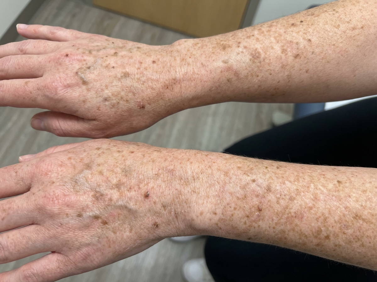
This patient presented with a defect. Which cellular process is most likely affected?
In PCR, why is Thermus aquaticus preferred over E. coli?
Which eukaryotic DNA polymerase is involved in proofreading and DNA repair during replication?
A young boy presents with difficulty in getting up from a sitting position and is diagnosed with Duchenne's muscular dystrophy. Which of the following statements is true regarding a mutation in the promoter region of the dystrophin gene?
An 8-year-old boy presents with progressive proximal muscle weakness and difficulty rising from the floor. Examination reveals calf pseudohypertrophy and a positive Gower's sign. What is the most common type of mutation in the gene responsible for this condition?
Aminoacyl-tRNA is required for which of the following processes?
Which technique is used to analyze DNA obtained from cancer biopsies when tumor cells are often contaminated with large numbers of admixed stromal cells?
Practice by Chapter
DNA Replication and Repair Mechanisms
Practice Questions
Transcription Factors and Gene Regulation
Practice Questions
Epigenetics and DNA Methylation
Practice Questions
RNA Processing and Splicing
Practice Questions
miRNA and RNA Interference
Practice Questions
Protein Synthesis and Post-Translational Modifications
Practice Questions
Genomics and Human Genome Project
Practice Questions
Single Nucleotide Polymorphisms
Practice Questions
Gene Therapy Approaches
Practice Questions
CRISPR-Cas9 and Genome Editing
Practice Questions
DNA Fingerprinting and Forensics
Practice Questions
Molecular Basis of Genetic Diseases
Practice Questions
Want unlimited practice?
Get full access to all questions, explanations, and performance tracking.
Scan to download app