
Molecular Biology and Genomics — MCQs

On this page
A frameshift mutation does not occur in multiples of which number?
All of the following are true regarding satellite DNA EXCEPT?
Which molecular method is used to locate a known gene locus?
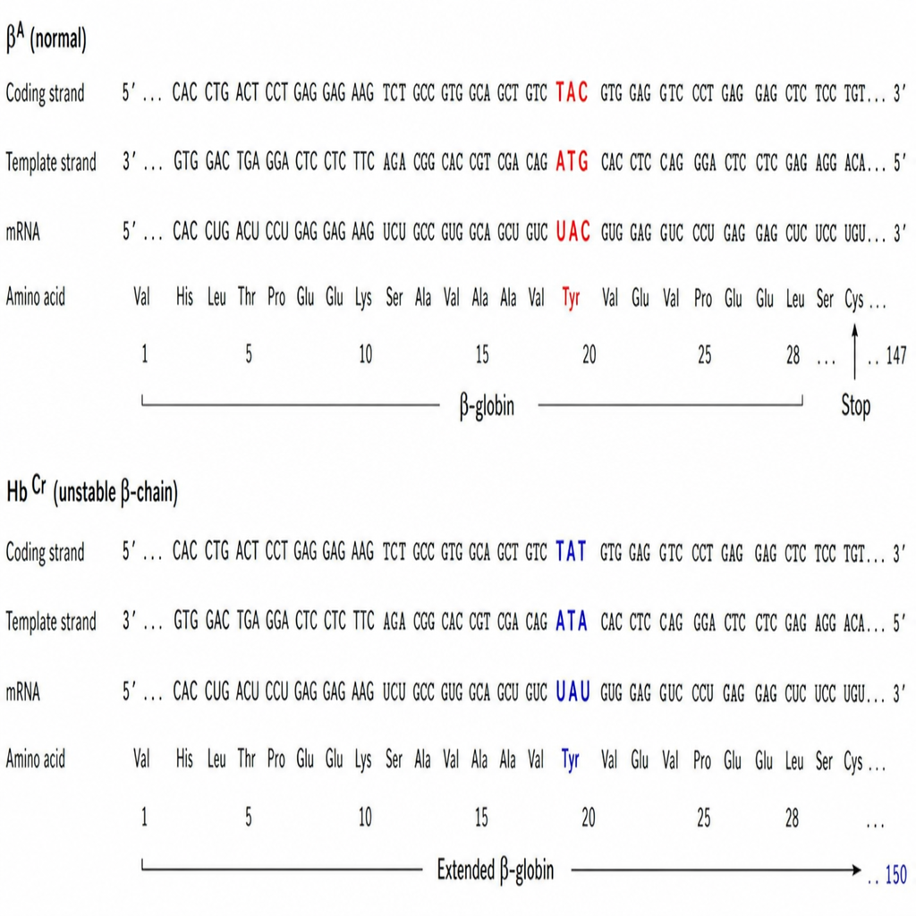
Hemoglobin electrophoresis of erythrocytes from a young child with anemia reveals an unstable hemoglobin, Hb Cranston (HbCr), with an abnormal b-globin chain. Given the nucleotide sequences of the normal b-globin gene (HbNl) and the HbCr b-chain, which of the following would account for the development of HbCr?
Which of the following is NOT required for a standard polymerase chain reaction?
What is the initial amino acid incorporated in prokaryotic protein synthesis?
Which of the following is an example of a non-coding RNA?
Which of the following cellular bodies is NOT found in the nucleus?
Chromosomal mutations can be identified by all of the following methods except:
Which of the following statements is true regarding Okazaki fragments?
Practice by Chapter
DNA Replication and Repair Mechanisms
Practice Questions
Transcription Factors and Gene Regulation
Practice Questions
Epigenetics and DNA Methylation
Practice Questions
RNA Processing and Splicing
Practice Questions
miRNA and RNA Interference
Practice Questions
Protein Synthesis and Post-Translational Modifications
Practice Questions
Genomics and Human Genome Project
Practice Questions
Single Nucleotide Polymorphisms
Practice Questions
Gene Therapy Approaches
Practice Questions
CRISPR-Cas9 and Genome Editing
Practice Questions
DNA Fingerprinting and Forensics
Practice Questions
Molecular Basis of Genetic Diseases
Practice Questions
Want unlimited practice?
Get full access to all questions, explanations, and performance tracking.
Scan to download app