
Molecular Biology and Genomics — MCQs

On this page
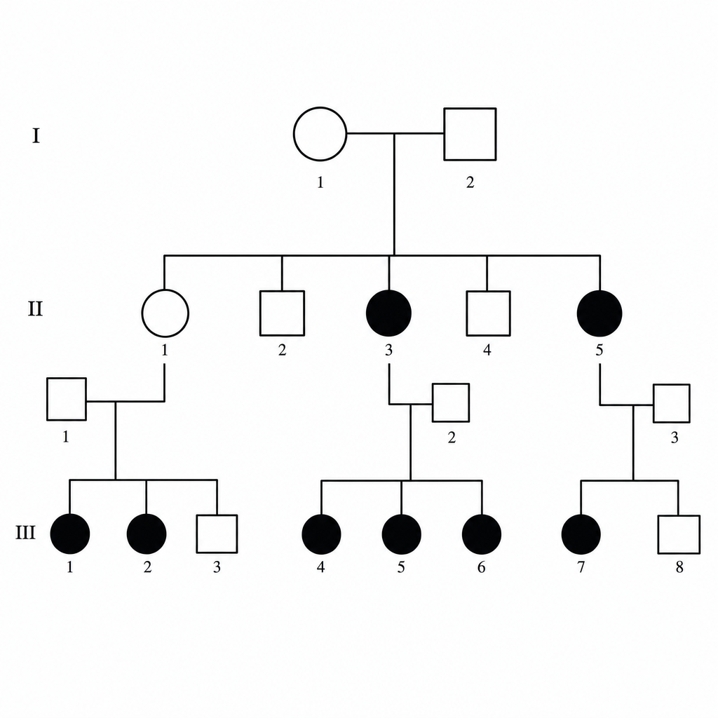
Analyze the following pedigree and give the mode of inheritance:
Which of the following enzymes is part of DNA dependent RNA polymerase?
Which of the following is a protein that is rich in basic amino acids and that functions in the packaging of DNA in chromosomes?
An 8-year-old boy presents with failure to thrive, alopecia totalis, localized scleroderma, a small face and jaw, a "beak" nose, wrinkled skin, and stiff joints. He has a single-point mutation in a nuclear protein that is silent in terms of the protein's primary structure. How could such a mutation lead to a disease?
Arrange the cyclins and CDKs in the cell cycle from G1 to S phase progression.
Prenatal diagnosis of Hemophilia is best done by?
Where does miRNA typically bind to facilitate gene knockdown?
Which DNA polymerase is/are involved in the repair of mammalian DNA?
Base substitution of GAC (Asp) to GAG (Glu) is an example of which type of mutation?
Which snRNA is not a part of the spliceosome?
Practice by Chapter
DNA Replication and Repair Mechanisms
Practice Questions
Transcription Factors and Gene Regulation
Practice Questions
Epigenetics and DNA Methylation
Practice Questions
RNA Processing and Splicing
Practice Questions
miRNA and RNA Interference
Practice Questions
Protein Synthesis and Post-Translational Modifications
Practice Questions
Genomics and Human Genome Project
Practice Questions
Single Nucleotide Polymorphisms
Practice Questions
Gene Therapy Approaches
Practice Questions
CRISPR-Cas9 and Genome Editing
Practice Questions
DNA Fingerprinting and Forensics
Practice Questions
Molecular Basis of Genetic Diseases
Practice Questions
Want unlimited practice?
Get full access to all questions, explanations, and performance tracking.
Scan to download app