
Molecular Biology and Genomics — MCQs

On this page
What is true about the coding strand of DNA?
What is an Okazaki fragment?
Which of the following is true about DNA polymerase in eukaryotes?
Senescent cells are deficient in what?
Aminoacyl t-RNA is required for all except?
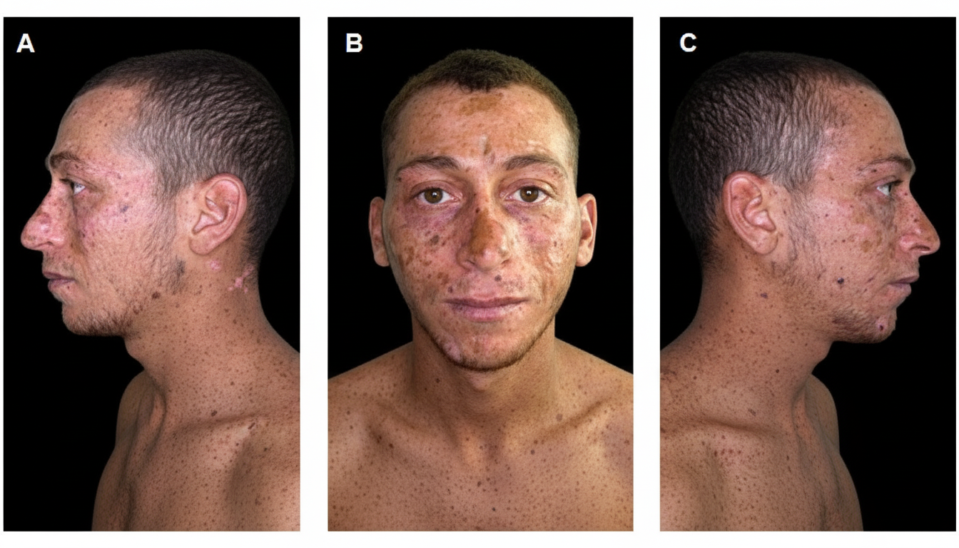
The disorder shown in the illustration is related to which DNA repair mechanism?
The attachment of eukaryotic mRNA to the ribosome is mediated through which of the following?
A mutation in a codon that causes a change in the coded amino acid is known as what?
All are steps of PCR EXCEPT?
Which cation is commonly used in PCR?
Practice by Chapter
DNA Replication and Repair Mechanisms
Practice Questions
Transcription Factors and Gene Regulation
Practice Questions
Epigenetics and DNA Methylation
Practice Questions
RNA Processing and Splicing
Practice Questions
miRNA and RNA Interference
Practice Questions
Protein Synthesis and Post-Translational Modifications
Practice Questions
Genomics and Human Genome Project
Practice Questions
Single Nucleotide Polymorphisms
Practice Questions
Gene Therapy Approaches
Practice Questions
CRISPR-Cas9 and Genome Editing
Practice Questions
DNA Fingerprinting and Forensics
Practice Questions
Molecular Basis of Genetic Diseases
Practice Questions
Want unlimited practice?
Get full access to all questions, explanations, and performance tracking.
Scan to download app