
Molecular Biology and Genomics — MCQs

On this page
In E. coli, the structural genes of the lac operon are stimulated under which condition?
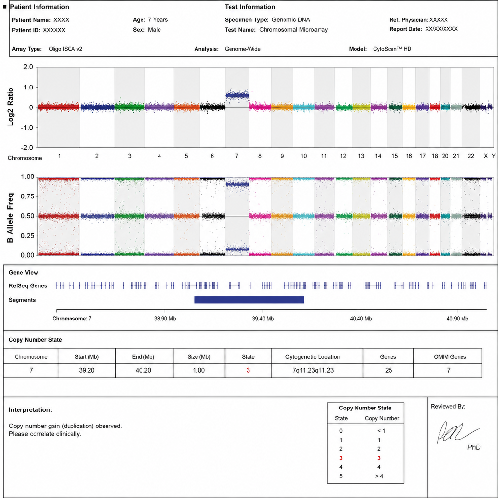
The test result of which investigation, for a child with developmental delay, is shown below?
Which of the following best describes the inheritance pattern of an X-linked recessive disease?
Which techniques are used to detect gene mutations?
All of the following require 5' capping except?
Which biomedical tool used in DNA technology utilizes an oligomer with a single base pair substitution?
Which of the following methods is most suited to assess the function of a gene?
Polypeptide chain termination is enhanced by:
Which of the following methods cannot be used to detect gene expression?
Which of the following is NOT a feature of the genetic code?
Practice by Chapter
DNA Replication and Repair Mechanisms
Practice Questions
Transcription Factors and Gene Regulation
Practice Questions
Epigenetics and DNA Methylation
Practice Questions
RNA Processing and Splicing
Practice Questions
miRNA and RNA Interference
Practice Questions
Protein Synthesis and Post-Translational Modifications
Practice Questions
Genomics and Human Genome Project
Practice Questions
Single Nucleotide Polymorphisms
Practice Questions
Gene Therapy Approaches
Practice Questions
CRISPR-Cas9 and Genome Editing
Practice Questions
DNA Fingerprinting and Forensics
Practice Questions
Molecular Basis of Genetic Diseases
Practice Questions
Want unlimited practice?
Get full access to all questions, explanations, and performance tracking.
Scan to download app