
Molecular Biology and Genomics — MCQs

On this page
What property of DNA synthesis does the Sanger method of DNA sequencing take advantage of to generate a sequencing ladder?
Genomic imprinting is seen in which of the following conditions?
Which of the following statements regarding satellite DNA is incorrect?
What is true about the coding strand of DNA?
Which of the following statements is NOT TRUE regarding the initiation of protein synthesis in eukaryotes?
Which of the following amino acids does not exhibit degeneracy in its genetic code?
How many base pairs are encoded by a prokaryotic polypeptide of 250 amino acids?
Restriction fragment length polymorphism is used for what purpose?
Co-translational insertion is seen with which of the following?
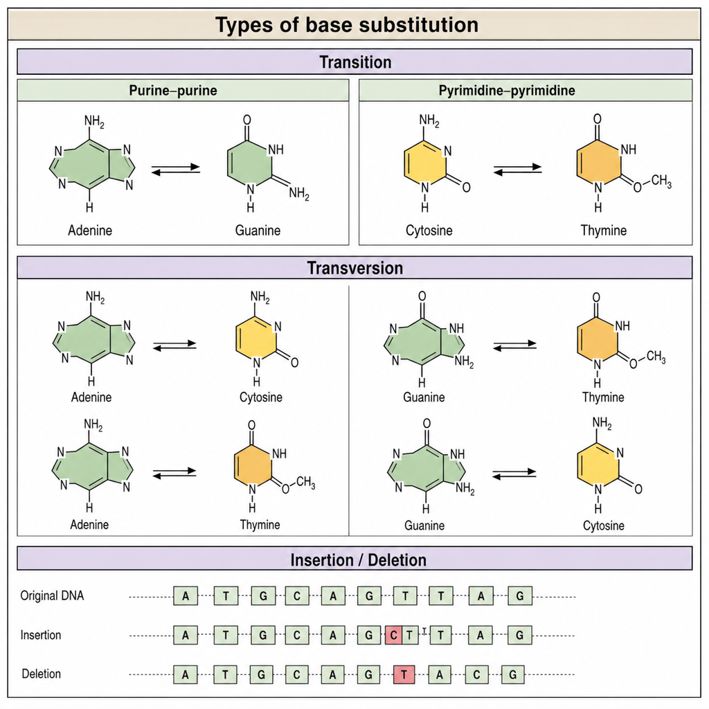
This is an example of which type of mutation?
Practice by Chapter
DNA Replication and Repair Mechanisms
Practice Questions
Transcription Factors and Gene Regulation
Practice Questions
Epigenetics and DNA Methylation
Practice Questions
RNA Processing and Splicing
Practice Questions
miRNA and RNA Interference
Practice Questions
Protein Synthesis and Post-Translational Modifications
Practice Questions
Genomics and Human Genome Project
Practice Questions
Single Nucleotide Polymorphisms
Practice Questions
Gene Therapy Approaches
Practice Questions
CRISPR-Cas9 and Genome Editing
Practice Questions
DNA Fingerprinting and Forensics
Practice Questions
Molecular Basis of Genetic Diseases
Practice Questions
Want unlimited practice?
Get full access to all questions, explanations, and performance tracking.
Scan to download app