
Molecular Biology and Genomics — MCQs

On this page
What banding technique is used for studying translocations involving the centromere?
Xeroderma pigmentosum is produced as a result of a defect in:
Which of the following is not an example of epigenetic change?
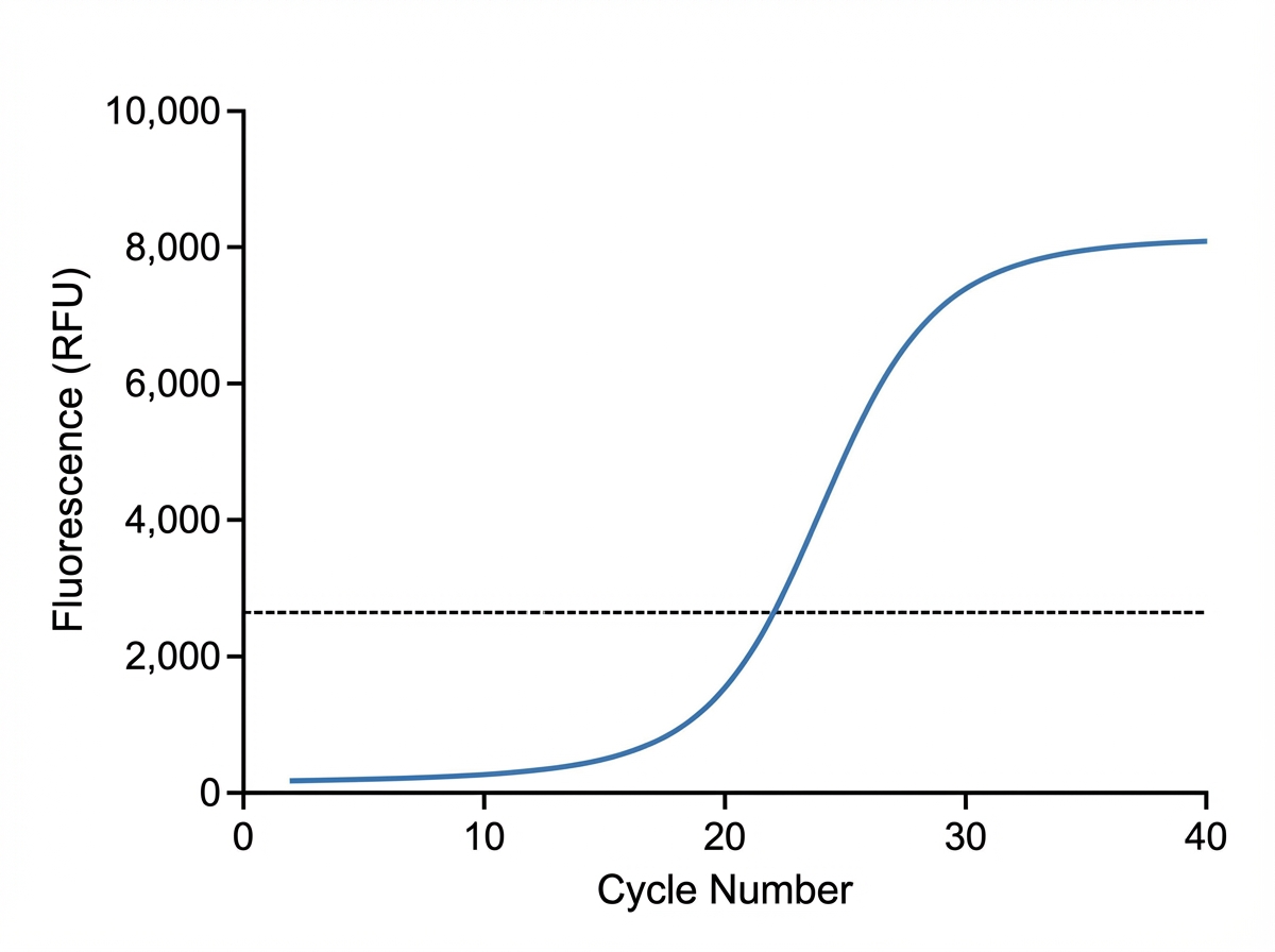
The graph below depicts a polymerase chain reaction. What type of PCR is shown?
Which codon is recognized by tRNA-Met?
What is the best method for assessing protein binding regions on a DNA molecule?
All of the following are DNA binding domains found in transcription factors, EXCEPT?
Which of the following is NOT a property of the Signal Recognition Particle (SRP)?
Which of the following statements are true about telomeres?
Introduction of DNA into cells with the help of electricity is known as?
Practice by Chapter
DNA Replication and Repair Mechanisms
Practice Questions
Transcription Factors and Gene Regulation
Practice Questions
Epigenetics and DNA Methylation
Practice Questions
RNA Processing and Splicing
Practice Questions
miRNA and RNA Interference
Practice Questions
Protein Synthesis and Post-Translational Modifications
Practice Questions
Genomics and Human Genome Project
Practice Questions
Single Nucleotide Polymorphisms
Practice Questions
Gene Therapy Approaches
Practice Questions
CRISPR-Cas9 and Genome Editing
Practice Questions
DNA Fingerprinting and Forensics
Practice Questions
Molecular Basis of Genetic Diseases
Practice Questions
Want unlimited practice?
Get full access to all questions, explanations, and performance tracking.
Scan to download app