
Molecular Biology and Genomics — MCQs

On this page
A change from UAC to UAG represents which type of mutation?
Which of the following statements regarding telomerase is true?
What is true about Polymerase Chain Reaction (PCR)?
Which of the following statements about microRNAs (miRNAs) is false?
All of the following are associated with changes in genetic material, except?
Which one of the following is the complementary sequence of 5' TTAAGCTAC 3'?
Which of the following statements about nucleotide excision repair is FALSE?
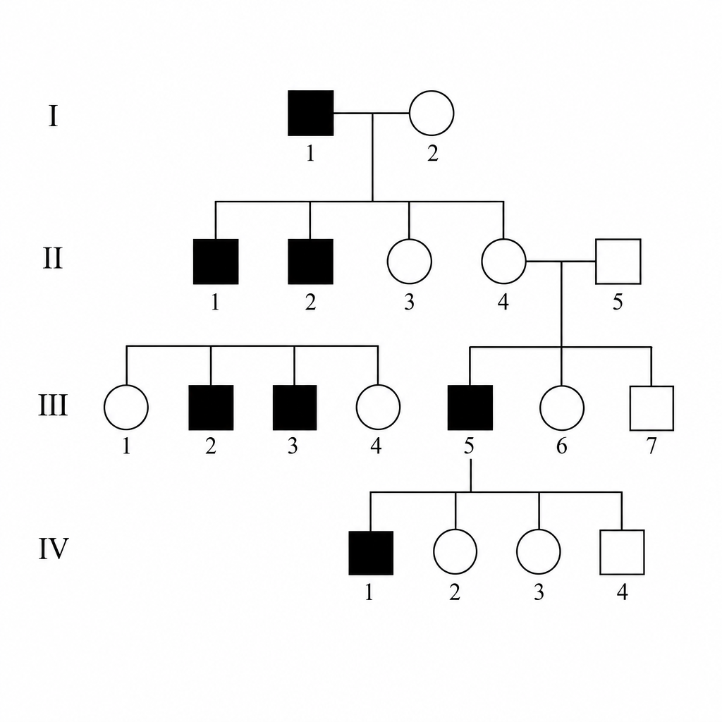
Examine the provided pedigree carefully. What type of transmission does it depict?
The Hershey-Chase experiment was performed on which of the following?
Which of the following activities is associated with 5' to 3' exonuclease activity?
Practice by Chapter
DNA Replication and Repair Mechanisms
Practice Questions
Transcription Factors and Gene Regulation
Practice Questions
Epigenetics and DNA Methylation
Practice Questions
RNA Processing and Splicing
Practice Questions
miRNA and RNA Interference
Practice Questions
Protein Synthesis and Post-Translational Modifications
Practice Questions
Genomics and Human Genome Project
Practice Questions
Single Nucleotide Polymorphisms
Practice Questions
Gene Therapy Approaches
Practice Questions
CRISPR-Cas9 and Genome Editing
Practice Questions
DNA Fingerprinting and Forensics
Practice Questions
Molecular Basis of Genetic Diseases
Practice Questions
Want unlimited practice?
Get full access to all questions, explanations, and performance tracking.
Scan to download app