
Carbohydrate Metabolism — MCQs

On this page
Which of the following is NOT a product of the uronic acid pathway in humans?
Which enzyme of glycolysis is also used in gluconeogenesis?
Which of the following is most effective for gluconeogenesis?
A patient presents with muscle cramps on exercise and positive myoglobinemia. What is the most likely disorder?
Conversion of lactate to glucose requires all except?
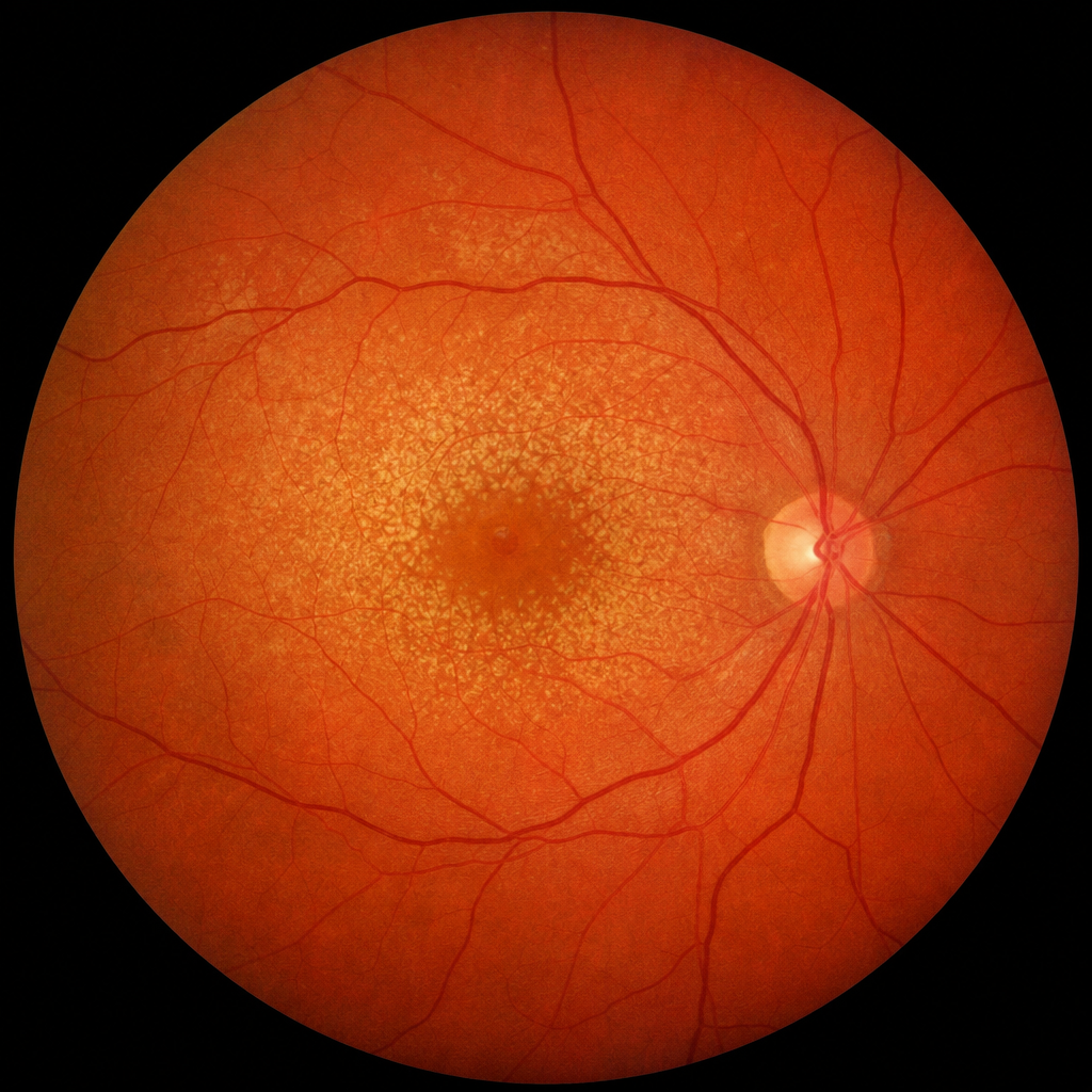
A newborn baby presents with a history of seizures and hepatomegaly. The following feature was noted on ophthalmic examination. What is the most probable diagnosis?
Which of the following types of reaction does NOT occur in glycolysis?
For which glycogen storage disease, enzyme replacement therapy is available?
What is the first product of glycogenolysis?
A 2-month-old infant presents with generalized muscle weakness and a "floppy infant" appearance. Additional features include macroglossia, feeding difficulty, hepatomegaly, and hypertrophic cardiomyopathy. What is the diagnosis?
Practice by Chapter
Carbohydrate Chemistry and Classification
Practice Questions
Glycolysis: Reactions and Regulation
Practice Questions
Gluconeogenesis: Reactions and Regulation
Practice Questions
Glycogen Metabolism: Synthesis and Breakdown
Practice Questions
Glycogen Storage Diseases
Practice Questions
Pentose Phosphate Pathway
Practice Questions
Metabolism of Fructose and Galactose
Practice Questions
Disorders of Fructose and Galactose Metabolism
Practice Questions
Blood Glucose Regulation
Practice Questions
Diabetes Mellitus: Biochemical Aspects
Practice Questions
Glycosylation and Glycoproteins
Practice Questions
Lactose Intolerance and Galactosemia
Practice Questions
Want unlimited practice?
Get full access to all questions, explanations, and performance tracking.
Scan to download app