Which particle size is most dangerous in causing pneumoconiosis?
The supraventricular crest is located between which of the following structures?
A 30-year-old man came with choking episodes after ingestion of fish bone while eating. The fishbone got impacted at the level of T4 in the esophagus. Which is the most likely site of obstruction?
While doing an endoscopy, constriction is felt at the oesophageal junction at 25 cm from the incisor. This is due to?
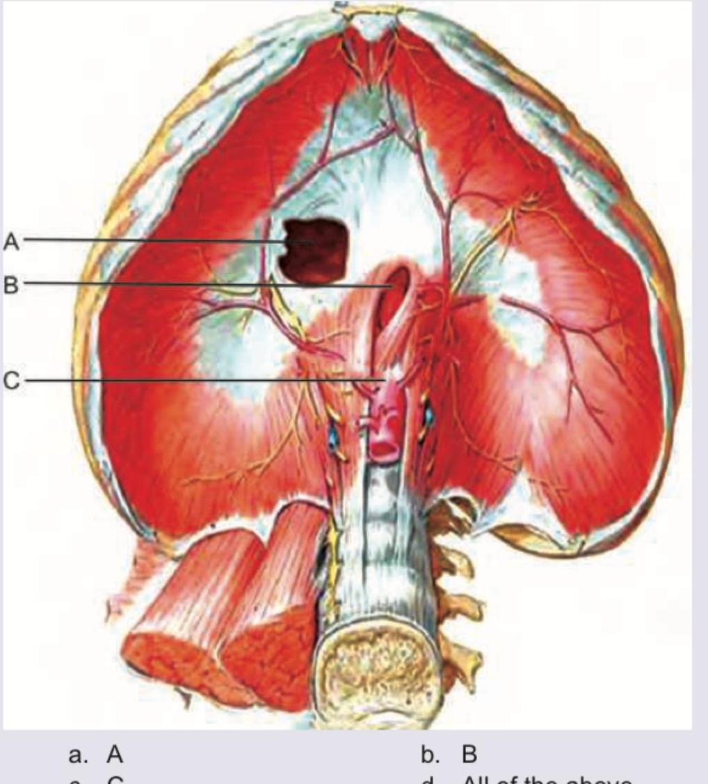
Which of the following structures passes through the foramen marked by the arrow? 
Match the cardiac anatomical features from Column I with their corresponding labels from Column II. Column I: 1. Receives oxygenated blood from pulmonary veins; 2. Lies anterior to ascending aorta; 3. Arises from LV; 4. Hypertrophy in pulmonary stenosis. Column II: A. Ascending aorta; B. Right ventricle; C. Pulmonary trunk; D. Left atrium.
The contraction of the diaphragm has no effect on which of the following apertures in the diaphragm?

The commonly seen depression deformity of the chest wall is known as
Inhaled foreign bodies are more likely to get lodged in the right main bronchus because
Consider the following statements: Venacaval opening of the diaphragm, situated at the level of T8 transmits 1. inferior vena cava 2. vagus nerve 3. branches of the right phrenic nerve 4. thoracic duct Which of the statements given above are correct?
Explanation: The pathogenicity of inhaled dust particles is primarily determined by their size, which dictates how deep they can travel into the respiratory tract and where they eventually settle. * **Why 1-5 microns is correct:** Particles in the **1 to 5-micron range** are the most dangerous because they are small enough to bypass the upper airway defenses (mucociliary escalator) but large enough to settle in the **terminal bronchioles and alveoli** via sedimentation. Once they reach the alveolar sacs, they are ingested by alveolar macrophages, triggering an inflammatory cascade and subsequent fibrosis (pneumoconiosis). **Analysis of Incorrect Options:** * **<1 micron (Option B):** These ultra-fine particles often behave like gas molecules; they remain suspended in the air and are typically **exhaled** back out without depositing in the lung parenchyma. * **5-15 micron (Option C):** Particles of this size are generally trapped by the mucus and cilia in the **tracheobronchial tree** and are cleared before reaching the gas-exchange zones. * **10-20 micron (Option D):** These large particles are filtered out by the **nasal vibrissae** (nose hairs) and the humidification process in the upper respiratory tract. **High-Yield Clinical Pearls for NEET-PG:** * **Silicosis:** The most common pneumoconiosis; characterized by "eggshell calcification" of hilar lymph nodes. * **Anthracosis:** Seen in coal miners; involves the accumulation of carbon pigment. * **Asbestosis:** Associated with "ferruginous bodies" (asbestos bodies) and increases the risk of bronchogenic carcinoma and mesothelioma. * **Key Concept:** The "Respiratory Zone" (where gas exchange occurs) begins at the respiratory bronchioles. Only particles <5 microns can effectively reach this zone to cause interstitial lung disease.
Explanation: The **supraventricular crest** (crista supraventricularis) is a thick muscular ridge located in the **right ventricle**. It serves as the anatomical boundary that separates the rough, inflow part of the ventricle from the smooth, outflow part (infundibulum/conus arteriosus). 1. **Why Option A is correct:** The right ventricle receives blood from the right atrium through the **tricuspid (atrioventricular) orifice** and ejects it through the **pulmonary orifice**. The supraventricular crest is positioned precisely between these two openings, arching over the anterior cusp of the tricuspid valve [1]. This separation helps streamline the flow of blood from the inflow tract to the outflow tract. 2. **Why other options are incorrect:** * **Option B:** The **fossa ovalis** is a feature of the interatrial septum in the **right atrium**, not the ventricle. * **Option C:** The junction between the superior vena cava and the right atrium is marked externally by the **sulcus terminalis** and internally by the **crista terminalis**. * **Option D:** The right and left coronary arteries arise from the **aortic sinuses** in the ascending aorta [1], unrelated to the internal muscular ridges of the right ventricle. **High-Yield Clinical Pearls for NEET-PG:** * **Inflow vs. Outflow:** The inflow part of the right ventricle is characterized by **trabeculae carneae**, while the outflow part (infundibulum) is smooth-walled. * **Moderator Band:** Also known as the **septomarginal trabecula**, it carries the right branch of the AV bundle and is another key landmark in the right ventricle. * **Embryology:** The smooth outflow tracts of both ventricles are derived from the **bulbus cordis**.
Explanation: ***Arch of aorta*** - The esophagus has three principal anatomic constrictions where foreign bodies, such as swallowed fish bones, commonly lodge; the second constriction occurs at the level of **T4/T5** where the **arch of the aorta** passes anteriorly. - This large anatomical structure compresses the esophagus against the vertebral column, creating a localized narrowing precisely matching the T4 level described for the obstruction [1]. *Left bronchus* - The **left main bronchus** also crosses anterior to the esophagus and contributes significantly to the formation of the **middle esophageal constriction** at the T4/T5 level. - However, in standard radiological and endoscopic visualization, the compression caused by the arch of the aorta is often considered the dominant landmark at this specific location [1]. *Cricopharyngeus* - This structure forms the initial and **uppermost esophageal constriction** (upper esophageal sphincter), located at the level of the **C6 vertebra** (cervical spine). - Obstruction at the cricopharyngeus would occur in the neck, which is anatomically much higher than the reported **T4 (thoracic spine)** impaction site. *Diaphragm* - The passage of the esophagus through the **esophageal hiatus** of the diaphragm forms the **third and lowest physiological constriction**, typically located at the vertebral level of **T10**. - This site of potential foreign body obstruction is significantly distal and caudal to the T4 level described in the clinical case. Management of such objects often involves careful endoscopy under general anesthesia to prevent perforation [2].
Explanation: ***b. Arch of aorta*** - The esophagus has **three normal anatomical constrictions** that are clinically important - At **25 cm from the incisors**, the esophagus is compressed by the **arch of aorta** crossing anteriorly, creating the **middle constriction** - This is the second of three constrictions and corresponds to the level of the **T4-T5 vertebrae** - These constrictions are sites where **foreign bodies may lodge** and where **strictures are more likely to develop** [1] *a. Inferior vena cava* - The IVC runs on the right side of the vertebral column and does **not cross anterior to the esophagus** - It does not cause any constriction of the esophagus *c. Right bronchus* - The **left main bronchus** (not right) crosses the esophagus anteriorly at approximately the same level as the aortic arch - The right bronchus does not come into contact with the esophagus *d. Diaphragmatic opening* - This causes the **third (lower) constriction** at approximately **40 cm from the incisors** - This is at the level of the **esophageal hiatus** in the diaphragm (T10 level) - Not at 25 cm as mentioned in the question
Explanation: ***Thoracic duct*** - The arrow in the image points to the **aortic hiatus**, which is the most posterior of the three major openings in the diaphragm, located at the vertebral level of **T12**. - This hiatus provides passage for the **aorta**, the **thoracic duct**, and the **azygos vein** from the thorax into the abdomen. *Oesophagus* - The oesophagus passes through its own opening, the **oesophageal hiatus**, which is located more anteriorly and superiorly at the **T10** vertebral level within the muscular part of the diaphragm. - This opening is formed by the fibers of the **right crus** of the diaphragm and also transmits the **vagal trunks**. *Inferior vena cava* - The inferior vena cava traverses the diaphragm through the **caval opening**, the most anterior and superior opening, situated at the **T8** vertebral level. - This foramen is located within the **central tendon** of the diaphragm and also allows passage for branches of the **right phrenic nerve**. *Sympathetic trunk* - The sympathetic trunk does not pass through any of the major apertures but rather descends posterior to the diaphragm. - It typically passes behind the **medial arcuate ligament** or pierces the **crus of the diaphragm** to enter the abdomen.
Explanation: 1-D, 2-C, 3-A, 4-B - 1-D (Receives oxygenated blood): The left atrium receives four pulmonary veins carrying oxygenated blood from the lungs [1], making it the only chamber that receives oxygenated blood directly from pulmonary circulation. - 2-C (Lies anterior to ascending aorta): The pulmonary trunk is positioned anteriorly and slightly to the left of the ascending aorta as both vessels exit from the base of the heart. - 3-A (Arises from LV): The ascending aorta arises directly from the left ventricle through the aortic valve and is the major systemic artery carrying oxygenated blood to the body. - 4-B (Hypertrophy in pulmonary stenosis): Pulmonary stenosis increases resistance to right ventricular outflow, causing chronic pressure overload and subsequent right ventricular hypertrophy. 1-A, 2-B, 3-C, 4-D - This matching is incorrect because the ascending aorta (A) does not receive blood from pulmonary veins [1]; it arises from the LV and carries blood away from the heart. Additionally, the pulmonary trunk (C) arises from the right ventricle, not the left ventricle. 1-B, 2-D, 3-C, 4-A - This matching is incorrect because the right ventricle (B) receives deoxygenated blood from the right atrium via the vena cavae, not oxygenated blood from pulmonary veins [1]. The left atrium (D) is a posterior chamber and does not lie anterior to the ascending aorta. 1-C, 2-A, 3-D, 4-B - This matching is incorrect because the pulmonary trunk (C) carries deoxygenated blood to the lungs; it does not receive oxygenated blood from pulmonary veins [1]. The left atrium (D) is not a vessel that arises from the LV—the correct structure is the ascending aorta (A).
Explanation: ***A*** - Label A points to the **vena cava foramen**, through which the inferior vena cava passes. This opening is located within the **central tendon** of the diaphragm. - The central tendon is **fibrous and non-contractile**, meaning that during diaphragm contraction, the vena cava foramen remains relatively unchanged in size. This ensures continuous venous return to the heart. *B* - Label B points to the **esophageal hiatus**, which transmits the esophagus and vagus nerves. - During diaphragm contraction, this opening **narrows slightly** due to the muscular action, aiding in the prevention of gastroesophageal reflux. *C* - Label C points to the **aortic hiatus**, through which the aorta, azygos vein, and thoracic duct pass. - While located behind the diaphragm, the muscular contraction of the crura surrounding the aortic hiatus can cause it to **change shape**, although it is less affected than the esophageal hiatus. *All of the above* - This option is incorrect because the contraction of the diaphragm **does affect** the esophageal and aortic hiatuses, albeit to varying degrees. - Only the vena cava foramen, located in the non-contractile central tendon, is largely unaffected.
Explanation: ***Pectus excavatum*** - This is a common congenital chest wall deformity characterized by an **inward depression of the sternum** and costal cartilages, creating a caved-in appearance of the chest. - It is often referred to as "funnel chest" due to the characteristic depression. *Kyphosis* - **Kyphosis** is an exaggerated, forward rounding of the back, often described as a hunchback. - It refers to a spinal curvature, not a depression of the chest wall itself. *Pectus carinatum* - **Pectus carinatum** is a chest wall deformity where the sternum protrudes outward, often referred to as "pigeon chest." - This is the opposite of a depression and involves a prominent, rather than sunken, chest. *Manubriosternal junction* - This term refers to the **normal anatomical landmark** where the manubrium meets the body of the sternum, also known as the sternal angle or angle of Louis. - It is not a deformity but a standard anatomical feature of the sternum.
Explanation: ***Right main bronchus is shorter, wider and nearly vertical*** - The **anatomical configuration** of the right main bronchus makes it a more direct continuation of the trachea. - Its **wider diameter** and **more vertical angle** allow foreign bodies to fall into it more easily due to gravity and airflow. *Left main bronchus is wider and shorter* - This statement is incorrect; the **left main bronchus** is actually **narrower and longer** than the right. - Its **more acute angle** stemming from the trachea also makes it less likely for foreign objects to lodge there directly. *Right main bronchus is shorter, narrower and lies horizontally in relation to trachea* - This statement is incorrect as the **right main bronchus** is indeed **shorter** but is **wider** and takes a **more vertical** course, not horizontal. - The description of being "narrower" and "horizontally" contradicts the anatomical characteristics that facilitate foreign body impaction. *Left main bronchus is narrow and longer* - This statement is anatomically correct for the left main bronchus; however, these characteristics make it **less likely** for foreign bodies to get lodged here. - The **narrower lumen** and **longer path**, combined with a more acute angle, make it a less direct route for inhaled objects.
Explanation: The **venacaval opening (caval hiatus)** is located at the level of **T8 vertebra** in the central tendon of the diaphragm. [1] **Statement 1: Inferior vena cava** ✓ - **CORRECT** - The inferior vena cava is the primary structure passing through the venacaval opening at T8. This opening is specifically designed to allow the IVC to pass from the abdomen into the thorax. **Statement 3: Branches of the right phrenic nerve** ✓ - **CORRECT** - The right phrenic nerve pierces the central tendon of the diaphragm near the venacaval opening. Some terminal branches of the right phrenic nerve pass through or adjacent to the caval opening to supply the inferior surface of the diaphragm [1]. *Statement 2: Vagus nerve* ✗ - *Incorrect* - The vagus nerves (anterior and posterior vagal trunks) pass through the **esophageal hiatus** at the level of **T10 vertebra**, not through the venacaval opening. [1] *Statement 4: Thoracic duct* ✗ - *Incorrect* - The thoracic duct passes through the **aortic hiatus** at the level of **T12 vertebra**, posterior to the diaphragm between the two crura. It does not pass through the venacaval opening. **Three major openings in the diaphragm:** - **T8** - Venacaval opening (IVC + right phrenic nerve branches) - **T10** - Esophageal hiatus (esophagus + vagus nerves) - **T12** - Aortic hiatus (aorta + thoracic duct + azygos vein) ***Therefore, statements 1 and 3 are correct.***
Thoracic Wall and Diaphragm
Practice Questions
Pleura and Lungs
Practice Questions
Mediastinum
Practice Questions
Heart and Pericardium
Practice Questions
Great Vessels and Azygos System
Practice Questions
Thoracic Duct and Lymphatics
Practice Questions
Autonomic Innervation
Practice Questions
Applied Anatomy and Clinical Correlations
Practice Questions
Thoracic Imaging and Cross-sectional Anatomy
Practice Questions
Embryological Development of Thoracic Structures
Practice Questions
Get full access to all questions, explanations, and performance tracking.
Start For Free