Which of the following vessels is typically injured in a patient with hemothorax?
The bronchial artery supplies all of the following except:
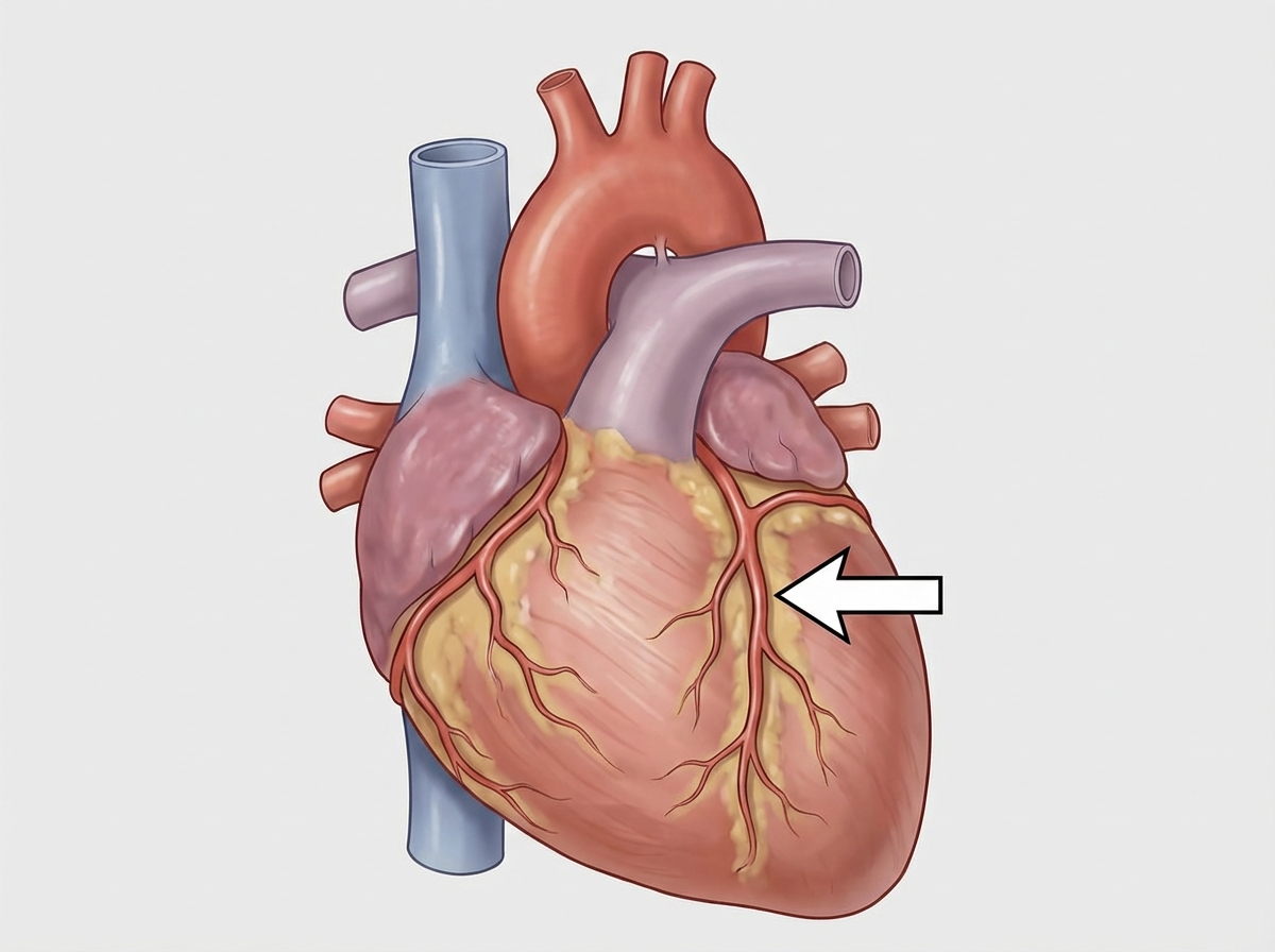
The structure marked with an arrow is:

Torus aorticus involves which structure?
Which of the following does NOT supply the oesophagus?
What is the root value of the intercostobrachial nerve?
Which muscle causes opening of the upper end of the esophagus?
Which nerve supplies the serratus anterior muscle?
A 35-year-old woman is admitted to the hospital with a complaint of shortness of breath. During physical examination, it is noted that there is wide splitting in her S2 heart sound. Which of the following valves is/are responsible for the production of the S2 heart sound?
Several weeks after surgical dissection of her left axilla for the removal of lymph nodes for staging and treatment of her breast cancer, a 32-year-old woman presented with "winging" of her left scapula when pushed against resistance. She also reported difficulty raising her right arm above her head. A nerve was accidentally injured during the surgical procedure, causing the scapular abnormality and inability to raise her arm normally. What was the origin of this injured nerve?
Explanation: Hemothorax is defined as the accumulation of blood in the pleural cavity, most commonly resulting from blunt or penetrating chest trauma. The **intercostal arteries** (branches of the thoracic aorta and internal thoracic artery) are the most frequent source of significant bleeding in these cases. Because these vessels are under **systemic arterial pressure**, injury to them leads to rapid accumulation of blood that does not easily tamponade, often requiring surgical intervention or chest tube drainage [1]. They are particularly vulnerable during procedures like thoracocentesis if the needle is not inserted correctly (along the superior border of the rib) [2]. **2. Why the Other Options are Incorrect:** * **Pulmonary Artery & Pulmonary Vein (A & B):** While injury to these major vessels causes massive hemothorax, they are located deep within the mediastinum and pulmonary hilum. They are less frequently injured in routine chest trauma compared to the peripheral chest wall vessels. Furthermore, the pulmonary circulation is a **low-pressure system**, which sometimes allows for spontaneous cessation of bleeding. * **Bronchial Artery (C):** These vessels supply the lung parenchyma and bronchi. While they are systemic vessels, they are rarely the primary source of a hemothorax; they are more commonly associated with **hemoptysis** (coughing up blood) in conditions like bronchiectasis or tuberculosis. **3. NEET-PG High-Yield Pearls:** * **Safe Zone for Thoracocentesis:** Always insert the needle at the **upper border of the rib** to avoid the neurovascular bundle (VAN: Vein, Artery, Nerve) located in the costal groove at the inferior border [2]. * **Internal Mammary (Thoracic) Artery:** Another common source of systemic bleeding in anterior chest wall trauma. * **Massive Hemothorax:** Defined as >1500 ml of blood or >200 ml/hr for 2–4 hours, usually indicating a systemic arterial bleed (like the intercostal artery) [1].
Explanation: The blood supply to the lungs is dual: the **bronchial circulation** (systemic) and the **pulmonary circulation**. The bronchial arteries arise from the descending thoracic aorta and supply the "conducting zone" of the respiratory system, providing oxygenated blood to the supporting tissues of the lungs [1]. **Why Alveolar Sac is the correct answer:** The bronchial arteries follow the bronchial tree only as far as the **respiratory bronchioles**. At this level, they anastomose with the pulmonary capillaries [1]. The structures involved in gas exchange—specifically the **alveolar ducts, atria, and alveolar sacs**—are supplied primarily by the **pulmonary circulation** (deoxygenated blood from the pulmonary artery). Therefore, the alveolar sac does not receive its primary supply from the bronchial artery. **Analysis of incorrect options:** * **A & B (Bronchus and Bronchioles):** The bronchial arteries provide nutrition to the walls of the bronchi and the conducting bronchioles up to the level of the terminal bronchioles [1]. * **C (Alveolar duct):** While the transition occurs at the respiratory bronchiole, most standard anatomical texts consider the bronchial artery's reach to terminate before the alveolar ducts and sacs, which are the functional units of the pulmonary circulation. **High-Yield Clinical Pearls for NEET-PG:** * **Venous Drainage:** Bronchial veins drain only the proximal portion of the lungs (near the hilum) into the **Azygos** (right) and **Hemiazygos** (left) veins [1]. The rest of the blood drains into the **Pulmonary veins**, creating a physiological right-to-left shunt. * **Origin:** Usually, there is **one** right bronchial artery (often arising from the 3rd posterior intercostal artery) and **two** left bronchial arteries (arising directly from the aorta). * **Clinical Significance:** In chronic inflammatory conditions like Bronchiectasis, bronchial arteries can hypertrophy, leading to massive **hemoptysis**.
Explanation: ***Coronary artery*** - The **coronary arteries** are visible on the **surface of the heart**, running in grooves (sulci) and supplying blood to the myocardium. - They originate from the **aortic root** just above the aortic valve and have a characteristic branching pattern across the heart's surface. *Pulmonary artery* - The **pulmonary trunk** and its branches are located **centrally** at the base of the heart, not on the surface. - It carries **deoxygenated blood** from the right ventricle to the lungs and appears as a large vessel emerging from the heart's base. *Arch of aorta* - The **aortic arch** is a large vessel that curves over the **superior aspect** of the heart and great vessels. - It gives rise to the **brachiocephalic trunk**, **left common carotid**, and **left subclavian arteries**, and is not typically visible on the heart's surface. *Pulmonary vein* - **Pulmonary veins** enter the **posterior aspect** of the left atrium and are not typically visible on the anterior surface. - They carry **oxygenated blood** from the lungs back to the heart and appear as smaller vessels entering the left atrium posteriorly.
Explanation: **Explanation:** The **Torus aorticus** is a distinct bulge or prominence found on the **septal wall of the right atrium**. It is produced by the pressure and anatomical proximity of the **ascending aorta** (specifically the right posterior or non-coronary sinus of Valsalva) as it lies immediately adjacent to the interatrial septum [1]. **Why Option D is Correct:** The right atrium’s medial wall is the interatrial septum. The aortic root is wedged between the two atria; however, its protrusion is most prominent into the anterosuperior portion of the right atrial septal wall, just above the limbus of the fossa ovalis. This elevation is the Torus aorticus. **Why Other Options are Incorrect:** * **Option A (Left Atrium):** While the aorta is related to the left atrium, it does not form a specific named "torus" there. The left atrium is primarily related to the esophagus and the descending aorta posteriorly. * **Option B & C (Arch and Ascending Aorta):** These are the structures *causing* the impression, not the structures *involved* by the name "Torus aorticus," which specifically refers to the atrial surface feature. **High-Yield Clinical Pearls for NEET-PG:** * **Surgical Landmark:** The Torus aorticus is a crucial landmark during transseptal catheterization and electrophysiological studies to avoid accidental puncture of the aortic root [1]. * **Triangle of Koch:** Located in the right atrium, its boundaries are the Tendon of Todaro, the septal leaflet of the tricuspid valve, and the orifice of the coronary sinus. It contains the **AV node**. * **Crista Terminalis:** A vertical ridge in the right atrium separating the smooth posterior part (sinus venarum) from the rough anterior part (pectinate muscles). The **SA node** is located at its upper end near the SVC opening.
Explanation: The esophagus is a muscular tube approximately 25 cm long, divided into three anatomical segments (cervical, thoracic, and abdominal). Its blood supply is segmental, derived from the nearest available major arteries. **Why Right Gastric Artery is the Correct Answer:** The **Right Gastric Artery** is a branch of the proper hepatic artery (or common hepatic) that supplies the lesser curvature of the **stomach**. It does not ascend high enough to contribute to the esophageal plexus. In contrast, the abdominal esophagus is primarily supplied by the **Left Gastric Artery** (a branch of the celiac trunk) and the left inferior phrenic artery. **Analysis of Incorrect Options:** * **A. Inferior Thyroid Artery:** This supplies the **cervical part** of the esophagus. It is a branch of the thyrocervical trunk. * **B. Descending Thoracic Aorta:** This supplies the **thoracic part** via direct esophageal branches. Additionally, bronchial arteries (branches of the aorta) also contribute to this segment. * **C. Left Gastric Artery:** This is the primary supply for the **abdominal part** of the esophagus. **High-Yield Clinical Pearls for NEET-PG:** * **Venous Drainage:** This is a critical site for **porto-systemic anastomosis**. The lower end drains into the Left Gastric Vein (Portal system) and the Azygos Vein (Systemic system). Obstruction leads to **esophageal varices**. * **Lymphatic Drainage:** Follows the arteries. Cervical → Deep cervical nodes; Thoracic → Posterior mediastinal nodes; Abdominal → Left gastric and celiac nodes. * **Constrictions:** Remember the four constrictions (at 15cm, 22cm, 27cm, and 40cm from incisors) as they are common sites for foreign body lodgment and corrosive injury.
Explanation: The **intercostobrachial nerve** is the lateral cutaneous branch of the **second intercostal nerve (T2)**. Unlike other intercostal nerves, its lateral branch does not divide into anterior and posterior branches. Instead, it pierces the external intercostal and serratus anterior muscles, crosses the axilla, and supplies the skin of the floor of the axilla and the upper medial aspect of the arm. [1] * **Option B (T2) is Correct:** The nerve originates specifically from the second thoracic spinal nerve. In some individuals, a small contribution from T3 may join it, but the primary root value is T2. * **Option A (T1):** The first intercostal nerve (T1) typically lacks a lateral cutaneous branch. Most of its fibers join the brachial plexus to supply the upper limb. * **Options C & D (T3 & T4):** These nerves give rise to standard lateral cutaneous branches that supply the skin of the chest wall in their respective dermatomes. While T3 can occasionally contribute to the intercostobrachial nerve, it is not the primary root. **Clinical Pearls for NEET-PG:** 1. **Referred Pain:** During a myocardial infarction (cardiac ischemia), pain is often referred to the inner aspect of the left arm. This occurs because visceral afferents from the heart and the intercostobrachial nerve both provide sensory input to the **T2 spinal segment**. 2. **Axillary Clearance:** This nerve is at high risk of injury during axillary lymph node dissection (e.g., in breast cancer surgery). [1] Damage results in numbness or paresthesia along the posteromedial aspect of the upper arm. 3. **Communication:** It often communicates with the medial cutaneous nerve of the arm.
Explanation: ### Explanation The **Cricopharyngeus** muscle is the correct answer because it functions as the **Upper Esophageal Sphincter (UES)**. **1. Why Cricopharyngeus is Correct:** The inferior constrictor muscle consists of two parts: the upper oblique fibers (**Thyropharyngeus**) and the lower horizontal fibers (**Cricopharyngeus**). Unlike other pharyngeal muscles that contract to propel food, the cricopharyngeus is **tonically contracted** at rest to prevent air from entering the esophagus and gastric contents from refluxing into the pharynx. During swallowing, this muscle **relaxes**, allowing the upper end of the esophagus to open and receive the food bolus [1]. **2. Why the other options are incorrect:** * **Epiglottis:** This is a cartilaginous structure, not a muscle. Its primary role is to flip downward to cover the laryngeal inlet during swallowing to prevent aspiration. * **Thyropharyngeus:** This is the upper part of the inferior constrictor. Its fibers are propulsive (peristaltic) in nature; it contracts to push food down rather than acting as a sphincter. * **Stylopharyngeus:** This is a longitudinal muscle of the pharynx (innervated by the Glossopharyngeal nerve). Its role is to **elevate** the larynx and pharynx during swallowing, not to open the esophageal lumen. **3. Clinical Pearls for NEET-PG:** * **Killian’s Dehiscence:** A potential gap exists between the thyropharyngeus and cricopharyngeus. This is a weak area where mucosal herniation can occur, leading to **Zenker’s Diverticulum**. * **Innervation:** While most pharyngeal muscles are supplied by the Pharyngeal Plexus (Vagus nerve), the **Stylopharyngeus** is the unique exception, supplied by the **Glossopharyngeal nerve (CN IX)**. * **Nerve Supply:** The cricopharyngeus receives motor supply from the **external laryngeal** and **recurrent laryngeal** nerves.
Explanation: The **Long thoracic nerve** (also known as the Nerve of Bell) is the correct answer. It arises from the ventral rami of the **C5, C6, and C7** nerve roots. It descends posterior to the brachial plexus and the first part of the axillary artery, running along the lateral surface of the serratus anterior muscle, which it supplies. The serratus anterior is crucial for protracting the scapula and rotating it upwards to allow for overhead abduction of the arm. **Analysis of Incorrect Options:** * **A. Thoracodorsal nerve (C6-C8):** Supplies the **Latissimus dorsi** muscle [1]. It is a branch of the posterior cord of the brachial plexus. * **B. Axillary nerve (C5-C6):** Supplies the **Deltoid** and **Teres minor** muscles. It passes through the quadrangular space. * **C. Musculocutaneous nerve (C5-C7):** Supplies the muscles of the **anterior compartment of the arm** (Biceps brachii, Coracobrachialis, and Brachialis). **Clinical Pearls for NEET-PG:** 1. **Winging of Scapula:** Damage to the long thoracic nerve (often during axillary lymph node dissection or radical mastectomy) leads to paralysis of the serratus anterior. This causes the medial border of the scapula to become prominent ("winging") when the patient pushes against a wall. 2. **Vulnerability:** Because the nerve runs superficially on the external surface of the muscle, it is highly susceptible to trauma or surgical injury. 3. **Mnemonic:** *"C5, 6, 7 raise your arms to heaven"* (referring to the nerve roots and the muscle's role in overhead abduction).
Explanation: The second heart sound (**S2**) is produced by the vibrations associated with the closure of the **semilunar valves** (Aortic and Pulmonary valves) at the end of ventricular systole [1]. **1. Why the Correct Answer is Right:** S2 marks the beginning of ventricular diastole. It consists of two components: **A2** (Aortic valve closure) and **P2** (Pulmonary valve closure) [1]. Under normal physiological conditions, A2 occurs slightly before P2 because the systemic resistance is higher than pulmonary resistance, causing the aortic valve to close earlier. During inspiration, the "splitting" of S2 becomes more pronounced as increased venous return to the right heart delays the closure of the pulmonary valve. **2. Analysis of Incorrect Options:** * **Option A & D (Mitral and Tricuspid valves):** These are **Atrioventricular (AV) valves**. Their closure at the beginning of ventricular systole produces the **First Heart Sound (S1)**. * **Option C (Aortic and Mitral valves):** This is incorrect because it mixes a semilunar valve (S2 component) with an AV valve (S1 component). **3. NEET-PG High-Yield Pearls:** * **Wide Splitting:** Seen in conditions that delay RV emptying (e.g., **Right Bundle Branch Block** or **Pulmonary Stenosis**). * **Fixed Splitting:** Pathognomonic for **Atrial Septal Defect (ASD)**. The split does not change with respiration. * **Paradoxical Splitting:** P2 occurs before A2; the split narrows during inspiration. Seen in **Left Bundle Branch Block** or **Aortic Stenosis**. * **A2 vs. P2:** A2 is normally louder and heard over the entire precordium, whereas P2 is softer and best heard at the left second intercostal space.
Explanation: **Explanation:** The clinical presentation of **"winging of the scapula"** following axillary surgery is a classic sign of injury to the **Long Thoracic Nerve**. This nerve innervates the **Serratus Anterior** muscle, which is responsible for protracting the scapula and holding its medial border against the thoracic wall. It also assists the Trapezius in rotating the scapula upward to allow for abduction of the arm above 90 degrees. **1. Why the Correct Answer is Right:** The Long Thoracic Nerve (Nerve of Bell) originates directly from the **Roots (ventral rami) of the brachial plexus**, specifically **C5, C6, and C7**. Because it arises so proximally and descends along the lateral thoracic wall, it is vulnerable during axillary lymph node dissection (ALND) or radical mastectomies [1]. **2. Why Incorrect Options are Wrong:** * **Option A (Upper Trunk):** The Suprascapular nerve and Nerve to Subclavius arise from the upper trunk, not the long thoracic nerve. * **Option B (Posterior Division):** Divisions of the plexus do not give off any branches; they merely redistribute fibers to the cords. * **Option D (Posterior Cord):** The posterior cord gives rise to the Axillary, Radial, Upper/Lower Subscapular, and Thoracodorsal nerves [1]. Injury to the Thoracodorsal nerve would cause weakness in extension and adduction (Latissimus dorsi), not winging. **Clinical Pearls for NEET-PG:** * **Mnemonic for Long Thoracic Nerve:** "C5, 6, 7 raise your arms to heaven." * **Winging Test:** Ask the patient to push against a wall; the medial border of the scapula becomes prominent (wings). * **Nerve at Risk:** During axillary surgery, two nerves are at high risk: the **Long Thoracic** (leading to winging) and the **Thoracodorsal** [1] (leading to "climbing" weakness/Latissimus dorsi palsy).
Thoracic Wall and Diaphragm
Practice Questions
Pleura and Lungs
Practice Questions
Mediastinum
Practice Questions
Heart and Pericardium
Practice Questions
Great Vessels and Azygos System
Practice Questions
Thoracic Duct and Lymphatics
Practice Questions
Autonomic Innervation
Practice Questions
Applied Anatomy and Clinical Correlations
Practice Questions
Thoracic Imaging and Cross-sectional Anatomy
Practice Questions
Embryological Development of Thoracic Structures
Practice Questions
Get full access to all questions, explanations, and performance tracking.
Start For Free