
Thorax — MCQs

On this page
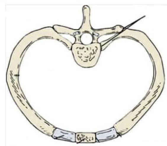
Identify the type of joint in the image provided.
Which of the following is not seen in the anterior mediastinum?
Which of the following statements about Sibson's fascia is correct?
Which of the following structures does not pass through the aortic opening?
What type of joint is formed by the costal cartilages of the 8th and 9th ribs?
Food can commonly get obstructed in the esophagus at all of the following locations except
Anterior Mediastinal nodes are included in which level of lymph nodes?
Which is the narrowest portion of the esophagus?
What is the average number of lobes in a human breast?
Which of the following does not directly drain into right atrium?
Practice by Chapter
Thoracic Wall and Diaphragm
Practice Questions
Pleura and Lungs
Practice Questions
Mediastinum
Practice Questions
Heart and Pericardium
Practice Questions
Great Vessels and Azygos System
Practice Questions
Thoracic Duct and Lymphatics
Practice Questions
Autonomic Innervation
Practice Questions
Applied Anatomy and Clinical Correlations
Practice Questions
Thoracic Imaging and Cross-sectional Anatomy
Practice Questions
Embryological Development of Thoracic Structures
Practice Questions
Want unlimited practice?
Get full access to all questions, explanations, and performance tracking.
Scan to download app