
Thorax — MCQs

On this page
A 30-year-old man came with choking episodes after ingestion of fish bone while eating. The fishbone got impacted at the level of T4 in the esophagus. Which is the most likely site of obstruction?
A woman presents with a breast lump, associated with skin dimpling and nipple retraction. What is the most likely anatomical structure responsible for the skin dimpling?
While doing an endoscopy, constriction is felt at the oesophageal junction at 25 cm from the incisor. This is due to?
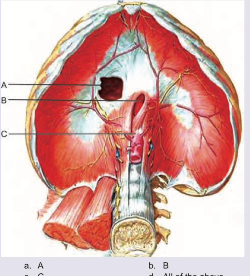
Which of the following structures passes through the foramen marked by the arrow? 
All of the following are superior mediastinal tumors except:
Match the cardiac anatomical features from Column I with their corresponding labels from Column II. Column I: 1. Receives oxygenated blood from pulmonary veins; 2. Lies anterior to ascending aorta; 3. Arises from LV; 4. Hypertrophy in pulmonary stenosis. Column II: A. Ascending aorta; B. Right ventricle; C. Pulmonary trunk; D. Left atrium.
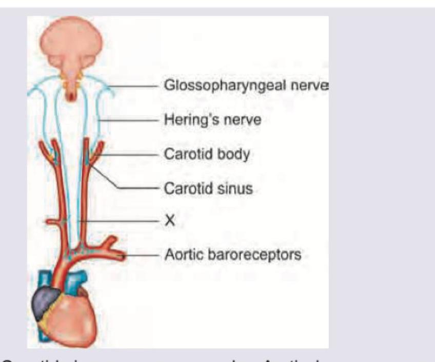
Which nerve marked as X innervates the Aortic Arch?
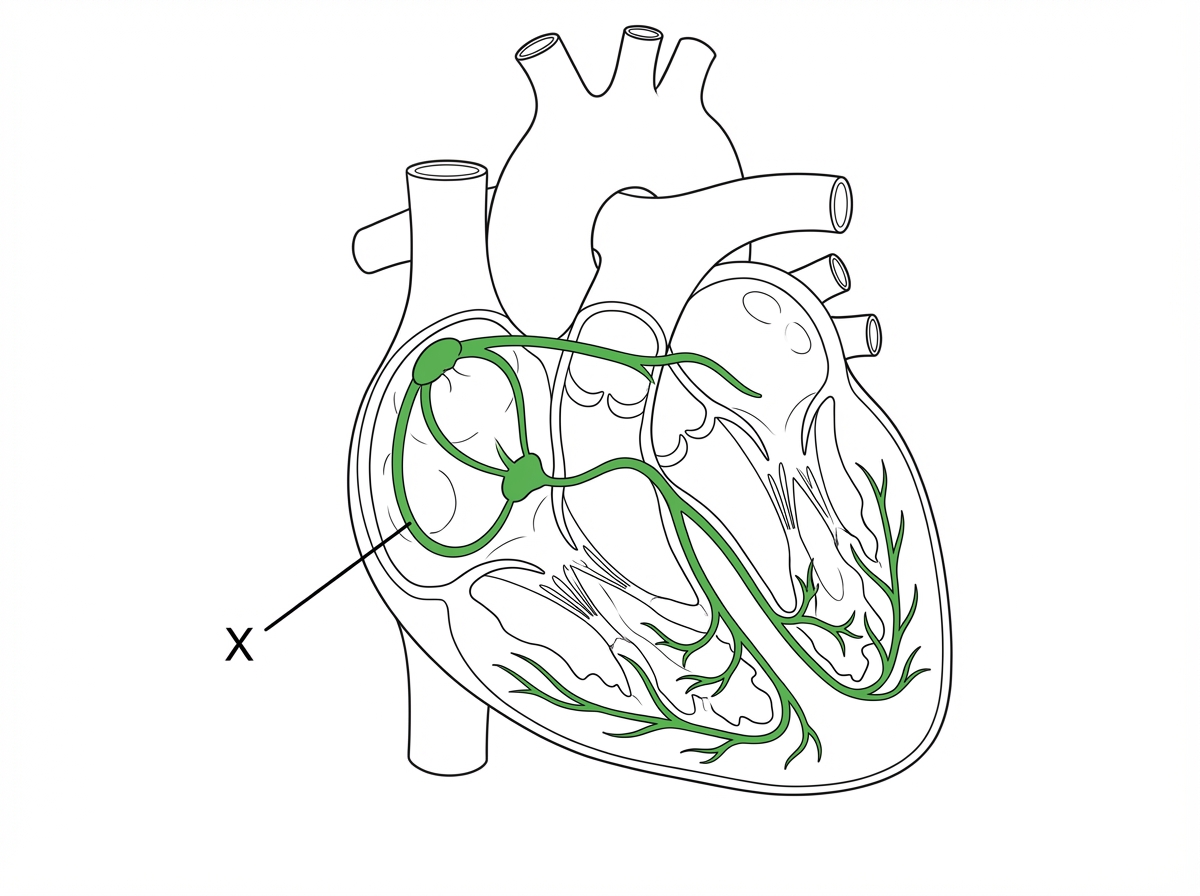
Identify the tract shown as X which is responsible for conduction from SA node to AV node.
The contraction of the diaphragm has no effect on which of the following apertures in the diaphragm?
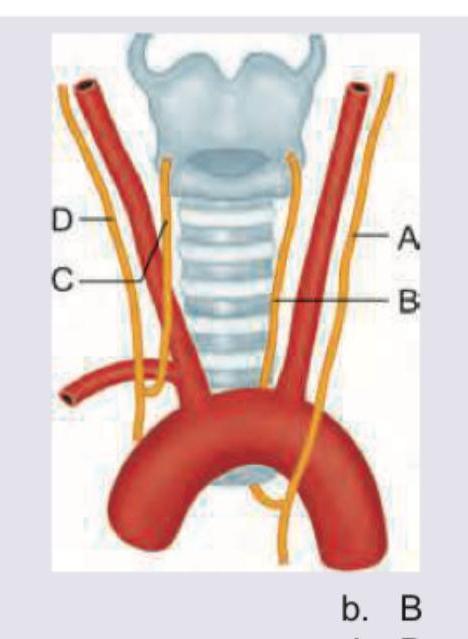
A 15-year-old child with rheumatic heart disease is having hoarseness of voice. Mark the nerve involved in the diagram shown below:
Practice by Chapter
Thoracic Wall and Diaphragm
Practice Questions
Pleura and Lungs
Practice Questions
Mediastinum
Practice Questions
Heart and Pericardium
Practice Questions
Great Vessels and Azygos System
Practice Questions
Thoracic Duct and Lymphatics
Practice Questions
Autonomic Innervation
Practice Questions
Applied Anatomy and Clinical Correlations
Practice Questions
Thoracic Imaging and Cross-sectional Anatomy
Practice Questions
Embryological Development of Thoracic Structures
Practice Questions
Want unlimited practice?
Get full access to all questions, explanations, and performance tracking.
Scan to download app