
Systemic Anatomy — MCQs

On this page
All of the following statements are true for the metaphysis of a bone except?
Which of the following is an example of a saddle joint?
All of the following are fibrous joints except?
Good collateral circulation occurs in which of the following structures?
All of the following organs have no lymphatic capillaries EXCEPT:
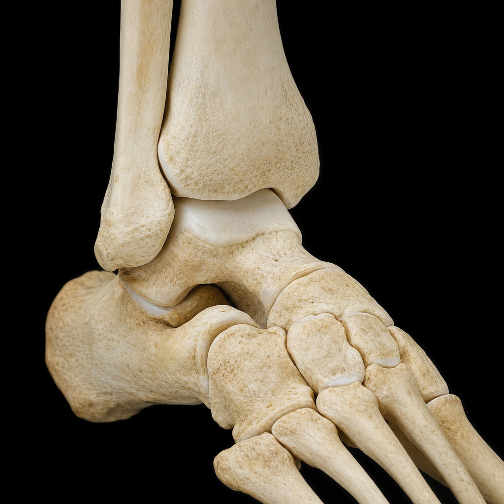
Identify the type of joint shown in the image.
Which of the following is NOT a spiral muscle?
Which artery supplies the growth plate of a long bone?
Which of the following is a traction epiphysis?
Which of the following is a discrete muscle of the Panniculus carnosus?
Practice by Chapter
Skeletal System
Practice Questions
Articular System
Practice Questions
Muscular System
Practice Questions
Cardiovascular System
Practice Questions
Lymphatic System
Practice Questions
Nervous System
Practice Questions
Respiratory System
Practice Questions
Digestive System
Practice Questions
Urinary System
Practice Questions
Reproductive System
Practice Questions
Endocrine System
Practice Questions
Integumentary System
Practice Questions
Want unlimited practice?
Get full access to all questions, explanations, and performance tracking.
Scan to download app