Identify the type of joint in the image provided.

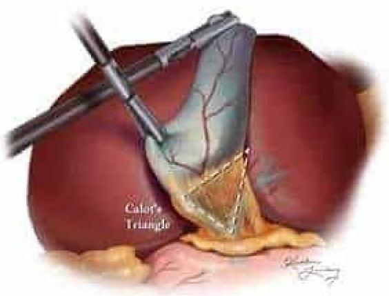
Which of the following structures is not a boundary of Calot's triangle shown in the given image?

What is the typical anatomical location of the parathyroid glands in relation to the thyroid gland?
Which part of the bone is considered the most vascular zone?
The narrowest part of the nasal cavity is?
Where are the stretch receptors located in the left atrium?
Which organ receives dual blood supply with both sources contributing to its primary metabolic function?
Seminal colliculus is present in ?
Which part of the stomach is primarily responsible for receiving and storing ingested food?
What is the correct sequence of uterine blood flow?
Explanation: ***Synovial joint*** - The image depicts a **costovertebral joint**, which connects a rib to a thoracic vertebra. These joints are **diarthrotic**, meaning they are freely movable, characteristic of synovial joints. - Synovial joints are characterized by the presence of a **synovial cavity**, articular cartilage, an articular capsule, and synovial fluid, allowing for a wide range of motion. *Syndesmosis* - A syndesmosis is a type of **fibrous joint** where two bones are joined by a ligament or a membrane, allowing for very limited movement, such as the distal tibiofibular joint. - This definition does not match the image, which shows a joint designed for movement between the rib and vertebra. *Synarthrosis* - Synarthrosis is a classification for **immovable joints**, such as sutures in the skull. - The costovertebral joints, as shown, allow for movement during respiration and are therefore not synarthrotic. *Symphysis* - A symphysis is a type of **cartilaginous joint** where bones are joined by **fibrocartilage**, allowing for slight movement. Examples include the pubic symphysis or intervertebral discs. - The costovertebral joint shown in the image is a synovial articulation, not a cartilaginous joint.
Explanation: ***Gallbladder*** - The image depicts **Calot's triangle**, which is an important anatomical landmark in gallbladder surgery. The gallbladder itself is located within this region, but it is not one of the defined boundaries of the triangle. - While central to the anatomy shown, the **gallbladder** is surrounded by the structures that form the triangle's boundaries rather than bounding it itself. *Common hepatic duct* - The **common hepatic duct** forms the medial boundary of Calot's triangle. - This duct is formed by the union of the right and left hepatic ducts and carries bile from the liver. *Cystic duct* - The **cystic duct** forms the lateral (or inferior) boundary of Calot's triangle. - This duct connects the gallbladder to the common hepatic duct. *Inferior surface of the liver* - The **inferior surface of the liver** forms the superior boundary of Calot's triangle. - Specifically, this refers to the edge of the right lobe of the liver at the base of the gallbladder fossa.
Explanation: ***Posterior aspect of the thyroid gland*** - The parathyroid glands are typically small, pea-sized endocrine glands located on the **posterior surface** of the thyroid gland [1] - There are usually **four parathyroid glands** (two superior and two inferior pairs) embedded within the thyroid capsule or closely apposed to its posterior lobes - This posterior positioning provides protection and maintains close anatomical relationship with the thyroid *Anterior aspect of the thyroid gland* - The anterior surface of the thyroid is covered by strap muscles (sternohyoid, sternothyroid) and skin [2] - Parathyroid glands are **not located anteriorly** as this would make them more vulnerable and less protected *Lateral aspect of the thyroid gland* - The lateral aspects of the thyroid lobes are adjacent to the carotid sheath structures - Parathyroid glands are not positioned laterally but remain on the **posterior surface** *Superior aspect of the thyroid gland* - While superior parathyroid glands are near the superior poles, their position is primarily **posterior**, not sitting directly on top [1] - The inferior parathyroid glands are located near the inferior poles, also on the posterior surface [1]
Explanation: ***Metaphysis*** - The metaphysis is the most **vascular** part of the bone, containing numerous blood vessels that supply the growing bone. - This high vascularity makes it a common site for **osteomyelitis** and bone tumors due to the abundant blood supply [1]. *Diaphysis* - The diaphysis is the **shaft** or central part of a long bone, primarily composed of **compact bone** [2]. - While it has blood supply through nutrient arteries, its vascularity is less dense compared to the metaphysis. *Epiphysis* - The epiphysis is the **end portion** of a long bone, typically covered by articular cartilage. - It receives its blood supply from periarticular vessels, but is less vascular than the metaphysis, especially in mature bone. *Medullary Cavity* - The medullary cavity is the central cavity of bone shafts where **bone marrow** is stored [2]. - While it contains hematopoietic stem cells and a rich blood supply, the surrounding bone tissue of the metaphysis itself is considered more vascular in terms of nutrient delivery and growth.
Explanation: ***Internal nasal valve*** - The **internal nasal valve** is considered the narrowest and most restrictive part of the **nasal airway**. - It is formed by the caudal border of the **upper lateral cartilage**, septum, and the head of the inferior turbinate. *Antrochoanal region* - The **antrochoanal region** is the posterior opening of the nasal cavity into the nasopharynx. - While it can be narrowed by polyps or mucosal swelling, it is not anatomically the **narrowest fixed point** of the nasal cavity. *1st nasal turbinate* - The **inferior turbinate** (often referred to as the 1st turbinate) can contribute to nasal resistance, especially when enlarged. - However, the turbinate itself is a structure that can vary in size and degree of congestion, but the **internal nasal valve** represents a consistently narrower anatomical choke point. *External nasal valve* - The **external nasal valve** is formed by the ala, columella, and nostril rim, and represents the entrance to the nasal cavity. - While it is a critical area for airflow, it is typically wider than the **internal nasal valve**.
Explanation: ***Entrance of the pulmonary veins*** - **Stretch receptors** are mechanoreceptors that detect changes in pressure and volume. In the left atrium, they are primarily located at the **junction of the pulmonary veins and the left atrium** [1]. - These receptors play a crucial role in the **Bainbridge reflex** and the release of **atrial natriuretic peptide (ANP)** in response to increased blood volume [1]. *Atrioventricular septum* - The **atrioventricular septum** separates the atria from the ventricles and primarily contains components of the **cardiac conduction system**, such as the AV node and bundle of His [2]. - While it has specialized tissues, it is not the primary location for **stretch receptors** involved in volume sensing. *Septum between the atria* - The **interatrial septum** primarily separates the right and left atria. - Although it contains some myocardial cells, it is not the main site for **stretch receptors** responsible for monitoring left atrial volume. *None of the options* - This option is incorrect because the **entrance of the pulmonary veins** is indeed the primary location for stretch receptors in the left atrium [1].
Explanation: ***Liver*** - The liver receives blood from two sources: the **hepatic artery** (supplying oxygenated blood, ~25% of blood flow) and the **hepatic portal vein** (supplying nutrient-rich, deoxygenated blood from the gastrointestinal tract, ~75% of blood flow). - Both blood supplies are essential for the liver's primary metabolic functions, detoxification, and nutrient processing [1]. - This is the classic example of dual blood supply in medical education. *Heart* - The heart receives its blood supply primarily from the **coronary arteries**, which branch off the aorta. - While it has an extensive arterial network, it has a single primary source of blood supply. *Kidney* - The kidneys receive their blood supply exclusively from the **renal arteries**, which branch directly from the aorta. - Each kidney typically has a single renal artery supplying it for high-pressure filtration. *Lung* - The lungs do receive blood from two sources: **pulmonary arteries** (deoxygenated blood for gas exchange) and **bronchial arteries** (oxygenated blood for tissue nourishment, <5% of flow). - However, the primary function (gas exchange) is served by pulmonary circulation alone, while bronchial circulation only nourishes lung tissue. - The liver is the standard answer for dual blood supply where both sources serve the organ's primary function.
Explanation: ***Correct: Urethra*** - The **seminal colliculus** (also known as the **verumontanum**) is a prominent ridge located on the posterior wall of the **prostatic urethra** - It contains the openings of the **ejaculatory ducts** and the **prostatic utricle** - This is a key anatomical landmark in the male urethra during endoscopic procedures *Incorrect: Prostate* - While the seminal colliculus is located within the portion of the urethra that passes through the prostate (prostatic urethra), it is not a structure *of* the prostate gland itself - The prostate is a gland that surrounds the urethra and contributes to seminal fluid - The seminal colliculus is an intraluminal urethral structure, not prostatic tissue *Incorrect: Testis* - The testis is the primary male reproductive organ responsible for **spermatogenesis** and hormone synthesis (testosterone) - It does not contain the seminal colliculus, which is located in the pelvic urethra *Incorrect: Scrotum* - The scrotum is an external dermal sac that houses the testes, epididymis, and lower spermatic cords - It provides temperature regulation for spermatogenesis - The seminal colliculus is an internal pelvic structure, not present in the scrotum
Explanation: ***Body*** - The **body (corpus)** is the largest part of the stomach, situated between the fundus and the antrum [1]. - This region is **primarily responsible for receiving and storing ingested food** as the main reservoir [2]. - It also produces **gastric acid and enzymes** (like pepsinogen) for the initial digestion of food [1]. - The body serves as the **principal storage chamber** where food accumulates after passing through the cardia [2]. *Fundus* - The **fundus** is the dome-shaped upper part of the stomach, located superior to the cardia. - While it can temporarily hold food and gas, it is **not the primary storage site** [2]. - Its main role is to serve as a **pressure buffer** and accumulation site for gases during digestion [2]. *Antrum* - The **antrum** is the lower, narrower part of the stomach, located before the pylorus. - It plays a crucial role in **mixing and grinding food with gastric juices** and propelling chyme towards the pylorus [1], [2]. - This is the **grinding chamber**, not a storage area [2]. *Pylorus* - The **pylorus** is the opening that connects the stomach to the duodenum, controlled by the pyloric sphincter. - Its main function is to **regulate the emptying of chyme** into the small intestine, not to store food [3].
Explanation: ***Uterine A → Arcuate A → Radial A → Spiral A*** - The **uterine artery** is the primary blood supply, branching into several arteries within the myometrium. - **Arcuate arteries** encircle the uterus, giving off perpendicular branches called **radial arteries**, which then give rise to the **spiral arteries** that supply the endometrium [1]. *Uterine A → Radial A → Arcuate A → Spiral A* - This sequence is incorrect because **radial arteries** arise from **arcuate arteries**, not the other way around. - **Arcuate arteries** are larger circumferential vessels that branch into the smaller radial arteries. *Uterine A → Spiral A → Radial A → Arcuate A* - This sequence is highly incorrect as **spiral arteries** are the most distal branches supplying the endometrium, not preceding radial or arcuate arteries [1]. - The flow is from larger to smaller vessels, with **spiral arteries** being the smallest and most terminal. *Uterine A → Arcuate A → Spiral A → Radial A* - This sequence incorrectly places **spiral arteries** before **radial arteries**. - **Radial arteries** are the immediate precursors to **spiral arteries**, providing direct branches to them.
Skeletal System
Practice Questions
Articular System
Practice Questions
Muscular System
Practice Questions
Cardiovascular System
Practice Questions
Lymphatic System
Practice Questions
Nervous System
Practice Questions
Respiratory System
Practice Questions
Digestive System
Practice Questions
Urinary System
Practice Questions
Reproductive System
Practice Questions
Endocrine System
Practice Questions
Integumentary System
Practice Questions
Get full access to all questions, explanations, and performance tracking.
Start For Free