
Systemic Anatomy — MCQs

On this page
Thoracic duct opens into ?
Lymphatic drainage of the thyroid gland is mainly?
Korner's septum is seen in?
Where is the scutum located in the middle ear?
Appendices epiploicae are a feature of?
Superficial epigastric artery is a branch of?
What is the typical number of lactiferous ducts that open in the nipple?
Olecranon process of ulna helps in formation of?
Which cervical vertebra has the longest spinous process?
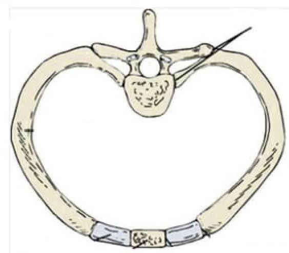
Identify the type of joint in the image provided.
Practice by Chapter
Skeletal System
Practice Questions
Articular System
Practice Questions
Muscular System
Practice Questions
Cardiovascular System
Practice Questions
Lymphatic System
Practice Questions
Nervous System
Practice Questions
Respiratory System
Practice Questions
Digestive System
Practice Questions
Urinary System
Practice Questions
Reproductive System
Practice Questions
Endocrine System
Practice Questions
Integumentary System
Practice Questions
Want unlimited practice?
Get full access to all questions, explanations, and performance tracking.
Scan to download app