Which of the following is true about the inferior vena cava (IVC)?
Which vein drains the superior pole of the thyroid gland?
During a dissection, a student notes a nerve lying on the serratus anterior muscle. Which nerve is it?
In an anatomy lab, students are asked to identify the structure that transports bile from the gallbladder to the common bile duct. What is this structure called?
What anatomical feature of the lymphatic system is primarily responsible for filtering pathogens from lymph?
Which cervical vertebra is known as the 'vertebra prominens' because of its distinctive spinous process?
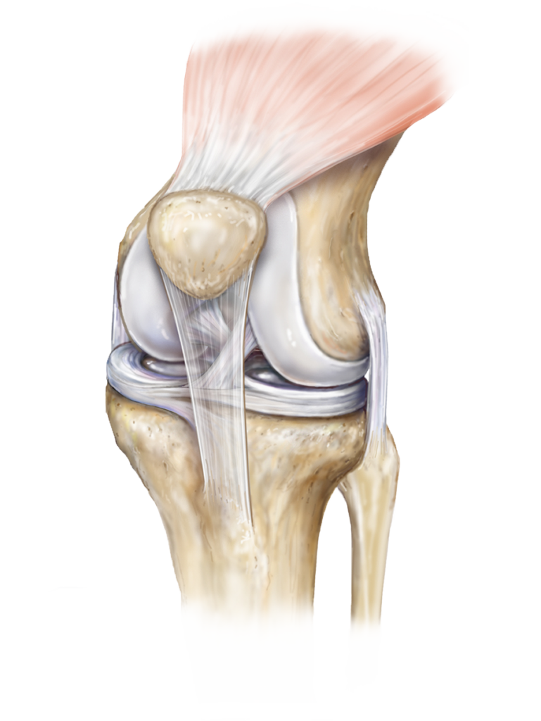
Identify the type of joint in the image provided.

Most mobile segment of vertebral column is -
Which of the following is the most metabolically active part of long bone?
Which of the following is not a part of the uveal tract?
Explanation: ***It drains blood from the lower body*** - The **inferior vena cava (IVC)** is a large vein that carries **deoxygenated blood** from the lower and middle body into the right atrium of the heart [1]. - It collects blood from veins such as the renal veins, hepatic veins, and iliac veins. *It receives blood from the pulmonary veins* - The **pulmonary veins** carry **oxygenated blood** from the lungs to the **left atrium** of the heart, not the IVC. - The IVC carries deoxygenated blood to the **right atrium** [1]. *It lies to the left of the aorta* - The **inferior vena cava** typically lies to the **right** of the aorta in the retroperitoneum. - The aorta is generally positioned more to the left of the midline compared to the IVC. *It passes through the aortic hiatus* - The **IVC** passes through its own opening in the diaphragm, the **caval opening**, at the level of T8. - The **aortic hiatus** is an opening in the diaphragm through which the **aorta** passes, located more posteriorly and inferiorly at T12.
Explanation: ***Superior thyroid vein*** - The **superior thyroid vein** specifically drains the **superior pole and anterior parts** of the thyroid gland. - It typically empties into the **internal jugular vein** and often accompanies the superior thyroid artery [1]. - Along with the middle and inferior thyroid veins, it forms part of the thyroid's venous drainage system. *Internal jugular vein* - While the internal jugular vein is a **major recipient** of thyroid venous drainage (receiving the superior and middle thyroid veins), it is not the drainage vessel directly from the gland itself. - It serves as a **main trunk** for several cervical and cranial veins. *Thyrocervical trunk* - The thyrocervical trunk is an **arterial branch** of the subclavian artery, not a venous structure. - It gives rise to the **inferior thyroid artery**, which supplies (not drains) the thyroid gland. - This is a common distractor testing knowledge of arterial vs. venous anatomy. *External jugular vein* - The external jugular vein primarily drains the **superficial structures of the head and neck**. - It does **not receive** direct drainage from the thyroid gland. - It empties into the subclavian vein.
Explanation: ***Long thoracic nerve*** - This nerve originates from the **brachial plexus** (C5-C7) and descends along the superficial surface of the **serratus anterior muscle**. - It innervates the **serratus anterior** and its injury leads to **winged scapula**. *Thoracodorsal nerve* - This nerve innervates the **latissimus dorsi muscle** and lies posterior to the axillary artery, not directly on the serratus anterior [1]. - It arises from the **posterior cord of the brachial plexus** (C6-C8). *Medial pectoral nerve* - This nerve penetrates the **pectoralis minor muscle** and innervates both the pectoralis major and minor [1]. - It runs on the deep surface of the pectoral muscles, not the serratus anterior [1]. *Lateral pectoral nerve* - This nerve innervates the **pectoralis major muscle** and pierces the clavipectoral fascia. - It typically runs lateral to the medial pectoral nerve and does not lie on the serratus anterior.
Explanation: ***Cystic duct*** - The **cystic duct** connects the **gallbladder** to the **common hepatic duct**, forming the **common bile duct** [1]. - Its primary function is to transport **bile** stored in the gallbladder both to and from the common bile duct [1]. *Hepatic duct* - The **common hepatic duct** is formed by the union of the **right and left hepatic ducts**, which drain bile from the liver. - It carries bile **from the liver** but does not directly transport bile *from the gallbladder* to the common bile duct. *Common bile duct* - The **common bile duct** is formed by the union of the **cystic duct** and the **common hepatic duct** [1]. - It transports bile to the duodenum but is not the direct channel *from the gallbladder* itself; the cystic duct serves that role [1]. *Pancreatic duct* - The **pancreatic duct** transports **pancreatic enzymes** and bicarbonate *from the pancreas* to the duodenum. - It plays no role in the transport of **bile** from the gallbladder.
Explanation: ***Lymph nodes*** - **Lymph nodes** are strategically located throughout the body to filter **lymph** and remove **pathogens**, cellular debris, and other foreign substances [1]. - They contain a high concentration of **immune cells**, such as **lymphocytes** and **macrophages**, which identify and destroy trapped foreign materials [2]. *Thymus* - The **thymus** is primarily involved in the **maturation and differentiation of T-lymphocytes**, not directly in filtering lymph from the general circulation. - It plays a crucial role in developing **immune tolerance** and is most active during childhood, undergoing atrophy in adulthood. *Spleen* - The **spleen** filters **blood**, not lymph, playing a significant role in removing old or damaged red blood cells and housing a large reservoir of immune cells [2]. - While it has immune functions, its primary role in filtration is within the bloodstream, not the lymphatic fluid. *Tonsils* - **Tonsils** are lymphoid tissues that form a protective ring in the pharynx, primarily acting as the first line of defense against **ingested or inhaled pathogens**. - They contain **lymphocytes** and help mount immune responses, but their filtration role is localized to the oral and pharyngeal cavities, not systemic lymph filtration.
Explanation: ***C7*** - The **C7 vertebra** is often called the **vertebra prominens** because its **spinous process** is typically the most prominent and easily palpable at the base of the neck. - This distinct feature makes it a crucial **anatomical landmark** for clinicians and therapists. *C1* - **C1** is known as the **atlas** and lacks a vertebral body and a spinous process, instead having an anterior and posterior arch. - Its primary role is to support the skull and facilitate head movements, notably nodding. *C2* - **C2** is known as the **axis** and is characterized by the **dens (odontoid process)**, which projects superiorly from its body, allowing for head rotation. - While it has a spinous process, it is usually bifid and not as prominent as that of C7. *C6* - The **spinous process of C6** is generally not as long or prominent as that of C7, although it is often bifid. - C6 is sometimes referred to as the "vertebrae prominens" in rare cases or in some anatomical variations, but C7 is the classic and most consistently prominent.
Explanation: ***Synovial joint*** - The image displays characteristics typical of a **synovial joint**, including **articular cartilage** covering bone ends, a **joint capsule**, visible **ligaments**, and a clearly defined joint space. - This type of joint allows for significant **movement** and is exemplified by the knee, which is shown in the image. *Syndesmosis joint* - A syndesmosis joint is a **fibrous joint** where bones are joined by a **ligament or interosseous membrane**, allowing for very little movement. - Examples include the **distal tibiofibular joint** or the **radioulnar syndesmosis**, which do not match the complex structure seen here. *Fibrous joint* - Fibrous joints are characterized by bones united by **fibrous connective tissue**, with sutures (e.g., in the skull) and gomphoses (teeth in sockets) being other examples. - They generally allow for **limited or no movement**, which is contrary to the highly mobile joint depicted. *Cartilaginous joint* - Cartilaginous joints are formed when bones are joined by **cartilage**, either **hyaline cartilage (synchondroses)** or **fibrocartilage (symphyses)**. - These joints allow for **some movement** (like the intervertebral discs or pubic symphysis) but lack the complex capsule, synovial fluid, and extensive range of motion seen in the image.
Explanation: ***Cervical*** - The **cervical spine** has the greatest range of motion due to the orientation of its **facet joints** and the relatively large intervertebral discs compared to vertebral body size. - This segment allows for extensive **flexion, extension, rotation, and lateral bending** of the head and neck. *Thoracic* - The **thoracic spine** has limited mobility primarily due to the attachment of the **rib cage**, which restricts movement. - The **facet joints** in this region are oriented to primarily allow for rotation, with less flexion/extension. *Lumbar* - The **lumbar spine** is designed for weight-bearing and stability, with its **facet joints** primarily allowing for flexion and extension. - **Rotation and lateral bending** are relatively limited compared to the cervical spine. *Sacral* - The **sacral spine** consists of five fused vertebrae, forming the sacrum, which articulates with the pelvic bones. - This fusion means the sacrum is virtually **immobile** at the segmental level and provides a stable base for the vertebral column.
Explanation: ***Metaphysis*** - The **metaphysis** is a highly vascular area between the **epiphysis** and **diaphysis** containing the **growth plate (physis)**. - It is characterized by active **bone remodeling** and formation, making it metabolically very active [1]. *Epiphysis* - The **epiphysis** is the end part of a **long bone**, where it articulates with other bones [1]. - While it contains **cancellous bone** and plays a role in bone health, its metabolic activity is generally lower than the metaphysis [1]. *Diaphysis* - The **diaphysis** is the **shaft** or central part of a **long bone**, primarily composed of compact bone [1]. - It is less metabolically active compared to the ends of the bone, primarily providing structural support. *Physis* - The **physis** (growth plate) is a cartilaginous disc located within the **metaphysis** responsible for bone lengthening [1]. - While extremely active in growth, the term 'metaphysis' encompasses the broader region of high metabolic activity, including the growth plate.
Explanation: ***Retina*** - The **retina** is the light-sensitive layer at the back of the eye, responsible for converting light into neural signals. It is part of the **sensory layer** of the eye, distinct from the uveal tract [1]. - While essential for vision, the retina originates from the **neural ectoderm** and is functionally separate from the uvea, which is primarily vascular and pigmented [2]. *Iris* - The **iris** is the colored part of the eye that surrounds the pupil and regulates the amount of light entering the eye. It is the **anterior-most part** of the uveal tract [3]. - It contains pigmented cells and smooth muscle fibers (sphincter and dilator pupillae) that control pupil size. *Ciliary body* - The **ciliary body** is a ring of tissue behind the iris that produces **aqueous humor** and contains the ciliary muscle, which is involved in accommodating the lens [3]. It is the **middle part** of the uveal tract. - It plays a crucial role in maintaining intraocular pressure and focusing vision [3]. *Choroid* - The **choroid** is the vascular layer of the eye, situated between the retina and the sclera, providing oxygen and nourishment to the outer layers of the retina [1]. It is the **posterior part** of the uveal tract. - Its rich blood supply and pigmentation help absorb excess light, preventing reflections within the eye.
Skeletal System
Practice Questions
Articular System
Practice Questions
Muscular System
Practice Questions
Cardiovascular System
Practice Questions
Lymphatic System
Practice Questions
Nervous System
Practice Questions
Respiratory System
Practice Questions
Digestive System
Practice Questions
Urinary System
Practice Questions
Reproductive System
Practice Questions
Endocrine System
Practice Questions
Integumentary System
Practice Questions
Get full access to all questions, explanations, and performance tracking.
Start For Free