All of the following are composite muscles, except:
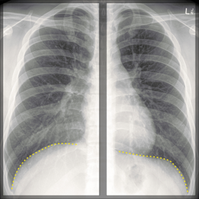
The ring is denoting the location of?

Absence of lymph nodes is characteristic of -
Bones are held by a long cord or sheet of dense fibrous connective tissue called:
Tubercles of Montgomery are present in
Circumference of an adult eyeball is:
The length of the female urethra is :
Which of the following is an end artery?
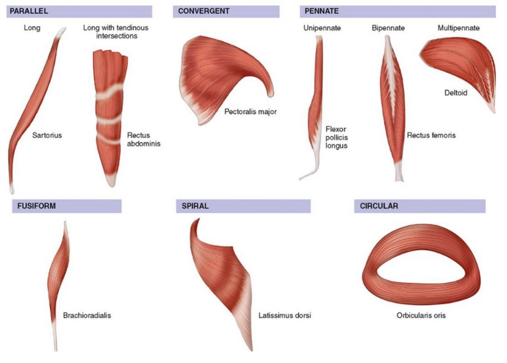
The muscle shown in the image is an example of which of the following type of muscle fiber arrangement?

Which is not a fibrous joint:
Explanation: ***Pectineus*** - The pectineus muscle is typically innervated by a **single nerve**, the **femoral nerve**, although it can sometimes receive a branch from the obturator nerve as well. - It is not characterized by distinct heads with different innervations, which is the defining feature of a composite muscle. - **This is the correct answer as pectineus is NOT a composite muscle.** *Flexor digitorum Profundus* - This is a **composite muscle** because its medial half is supplied by the **ulnar nerve**, and its lateral half is supplied by the **median nerve** (anterior interosseous branch). - Its different parts arise embryologically from separate muscle masses, leading to dual innervation. *Biceps Femoris* - The biceps femoris is a **composite muscle** as its **long head** is innervated by the **tibial division** of the sciatic nerve, while its **short head** is innervated by the **common fibular (peroneal) division** of the sciatic nerve. - This dual innervation reflects its development from two distinct muscle precursors. *Adductor Magnus* - The adductor magnus is a **composite muscle** with dual innervation: its **adductor portion** (upper fibers) is supplied by the **obturator nerve**, while its **hamstring portion** (lower fibers forming the ischiocondylar part) is supplied by the **tibial division** of the sciatic nerve. - This reflects its embryological origin from both adductor and hamstring muscle masses.
Explanation: ***Tricuspid valve*** - The image likely displays a **cardiac outline** on an X-ray, and the ring points to the typical location of the **tricuspid valve** within the right side of the heart. - The tricuspid valve separates the **right atrium** from the **right ventricle**. *Pulmonary valve* - The pulmonary valve is located anterior and to the left of the tricuspid valve, separating the **right ventricle** from the **pulmonary artery**. - Its position is generally superior and more towards the midline compared to the ring's location. *Mitral valve* - The mitral valve is situated on the **left side of the heart**, between the **left atrium** and **left ventricle**. - Its location is distinctly different from the highlighted area. *Aortic valve* - The aortic valve is located between the **left ventricle** and the **aorta**, typically found superior and central in relation to the cardiac silhouette. - This position is away from the area indicated by the ring.
Explanation: ***Brain*** - The **central nervous system (CNS)**, including the brain, is the **classic textbook answer** for organs lacking lymph nodes due to its status as an immunologically privileged site [1]. - The **blood-brain barrier** prevents conventional lymphatic drainage, and there are **no organized lymph nodes** within the brain parenchyma [1]. - Recent evidence suggests lymphatic-like drainage pathways (glymphatic system and meningeal lymphatics) that clear waste, but these do **not contain lymph nodes**. - Brain is the **most definitive answer** among organs completely devoid of lymph nodes. *Placenta* - The placenta is also an immunologically privileged organ that **lacks organized lymph nodes**. - However, it is a **temporary organ** (present only during pregnancy), whereas the brain is a permanent organ, making brain the more classical answer in exam contexts. - Its immune regulation occurs through specialized cellular mechanisms rather than lymph node-mediated immunity. *Lung* - The lungs are rich in **lymphatic vessels** and contain numerous **lymph nodes**, particularly in the hilar and mediastinal regions [2], [3]. - These lymph nodes play a crucial role in immune surveillance and drainage of lymphatic fluid from the pulmonary tissues. - **Clearly has lymph nodes** - incorrect option. *Liver* - The liver has an extensive **lymphatic system** and contains regional lymph nodes, primarily located in the **porta hepatis**. - These lymph nodes contribute to immune responses against pathogens entering via the portal circulation. - **Clearly has lymph nodes** - incorrect option.
Explanation: ***Syndesmosis*** - A **syndesmosis** is a type of fibrous joint where bones are joined by a cord or sheet of **dense fibrous connective tissue**, such as an interosseous membrane. - Examples include the articulation between the **tibia and fibula**, and the **ulna and radius**, allowing for limited movement. *Synchondrosis* - A **synchondrosis** is a cartilaginous joint joined by **hyaline cartilage**, typically seen in temporary joints like epiphyseal plates. - This type of joint eventually **ossifies** in adulthood, forming a synostosis. *Gomphosis* - A **gomphosis** is a fibrous joint that anchors a tooth to its bony socket, characterized by the insertion of a **peg-like structure** into a socket. - The connective tissue in a gomphosis is primarily the **periodontal ligament**. *Suture* - A **suture** is a type of fibrous joint found only in the skull, connecting bones with a narrow, interlocking seam of **dense fibrous connective tissue** [1]. - These joints are **immobile** in adults, providing protection for the brain.
Explanation: ***Breast*** - **Tubercles of Montgomery** are sebaceous glands found on the **areola**, the pigmented area surrounding the nipple [1]. - They become more prominent during pregnancy and lactation, producing an oily secretion that helps lubricate and protect the nipple and areola [1]. *Lung* - The **lungs** are responsible for gas exchange and contain structures like bronchi, bronchioles, and alveoli. - There are no anatomical structures within the lung parenchyma or airways referred to as Tubercles of Montgomery. *Duodenum* - The **duodenum** is the first part of the small intestine, primarily involved in digestion and absorption. - Its inner surface is characterized by plicae circularis, villi, and crypts, none of which are known as Tubercles of Montgomery. *Liver* - The **liver** is a large organ with diverse metabolic functions, composed of hepatocytes arranged in lobules. - While it has a complex anatomical structure, it does not contain any Tubercles of Montgomery.
Explanation: ***75 mm*** - The mean **circumference of an adult human eyeball** is approximately 75 mm. - This corresponds to an approximate diameter of 24 mm. *70 mm* - This value is slightly less than the average circumference of an adult eyeball. - While dimensions can vary, 70 mm is generally considered smaller than the typical range. *65 mm* - This circumference is significantly smaller than the average adult eyeball and would be considered an abnormally small globe, known as **microphthalmia**. - Such a size would likely be associated with visual impairment. *80 mm* - This value represents a circumference larger than the average adult eyeball and would correspond to an abnormally large globe, potentially indicative of conditions like **buphthalmos** (enlargement due to high intraocular pressure). - This increased size can affect ocular function and refraction.
Explanation: ***40 mm*** - The typical length of the **female urethra** is approximately **40 mm** (or 4 cm). - This relatively short length is a key reason why females are more susceptible to **urinary tract infections** compared to males. *20 mm* - **20 mm** (2 cm) is significantly shorter than the average female urethral length. - Such a short urethra would drastically increase the risk of **ascending infections** into the bladder. *60 mm* - **60 mm** (6 cm) is longer than the typical female urethra. - A urethra of this length would be more comparable to conditions in males or specific anatomical variations, not the average female. *45 mm* - While closer to the correct answer, **45 mm** is still slightly longer than the generally accepted average length for the female urethra. - The textbook average is usually cited as **4 cm** or 40 mm.
Explanation: ***Interlobar artery*** - The **interlobar arteries** are true **end arteries** of the kidney with **no anastomoses** between adjacent vessels. - They arise from segmental arteries and run between the renal pyramids toward the corticomedullary junction. - Occlusion of an interlobar artery leads to **segmental infarction** of the kidney tissue it supplies, as there is no collateral circulation. - This is clinically important in cases of renal embolism or thrombosis. *Arcuate artery* - **Arcuate arteries** branch from interlobar arteries and run along the base of the renal pyramids at the corticomedullary junction. - These arteries form **incomplete arches** and have **anastomotic connections** with adjacent arcuate arteries. - Due to these anastomoses, they are **not considered true end arteries**. *Interlobular artery* - **Interlobular arteries** (also called cortical radial arteries) arise from arcuate arteries and extend into the renal cortex. - They give off afferent arterioles to supply glomeruli. - While they have minimal collateral flow, some sources classify them as functional end arteries, but the **interlobar arteries** are the classic example. *Renal artery* - The **renal artery** is the main arterial supply to the kidney and divides into **segmental arteries**. - It is not an end artery as it has multiple large branches that can provide collateral circulation. - The segmental branches further divide into interlobar arteries, which are the actual end arteries.
Explanation: ***Convergent*** - The image clearly depicts the **Pectoralis major** muscle, which is a classic example of a **convergent muscle** (also known as triangular muscles). - **Convergent muscles** have a broad origin and converge to a single, narrow insertion, allowing for varied actions depending on which portion of the muscle contracts. *Bipennate* - **Bipennate muscles** have muscle fibers that are arranged on both sides of a central tendon, resembling a feather. - Examples include the **Rectus femoris**. This morphology is not depicted in the question's image for the *Pectoralis major*. *Cruciate* - The term **cruciate** describes muscles or ligaments that cross each other in the shape of an "X." - This is not a standard categorization for muscle fiber arrangement as shown in the provided muscle types. *Spiral* - **Spiral muscles** have fibers that twist as they run from origin to insertion, such as the **Latissimus dorsi**. - The muscle in the image (Pectoralis major) does not exhibit this twisting characteristic.
Explanation: ***First costochondral joint*** - This is a **cartilaginous joint (synchondrosis)**, where the **first rib connects to its costal cartilage**. - It is classified as a **primary cartilaginous joint** and is immovable. - Unlike other costochondral joints, the first is a true synchondrosis with hyaline cartilage. *Gomphoses* - These are **fibrous joints** that anchor the teeth into the alveolar sockets of the jawbone. - The connection is made by the **periodontal ligament**, composed of dense fibrous connective tissue. *Cranial sutures* - These are **fibrous joints** found only in the skull, connecting the cranial bones. - They are designed for **minimal to no movement** to protect the brain. *Inferior tibiofibular joint* - This is a **fibrous joint known as a syndesmosis**, where the tibia and fibula are united by a strong interosseous ligament. - It allows for **limited movement** and provides stability to the ankle joint.
Skeletal System
Practice Questions
Articular System
Practice Questions
Muscular System
Practice Questions
Cardiovascular System
Practice Questions
Lymphatic System
Practice Questions
Nervous System
Practice Questions
Respiratory System
Practice Questions
Digestive System
Practice Questions
Urinary System
Practice Questions
Reproductive System
Practice Questions
Endocrine System
Practice Questions
Integumentary System
Practice Questions
Get full access to all questions, explanations, and performance tracking.
Start For Free