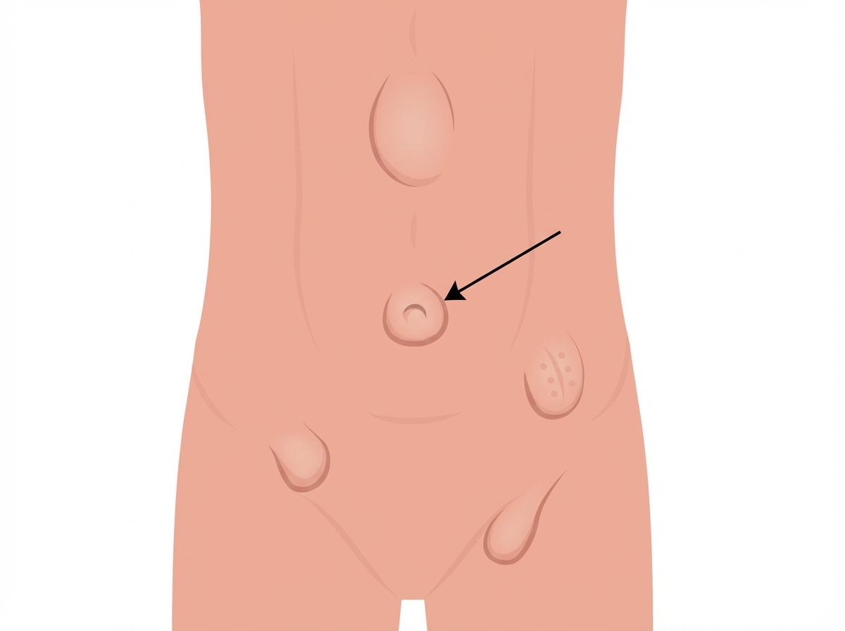
The dermatome level of the structure indicated in the image is: (Recent NEET Pattern 2019)

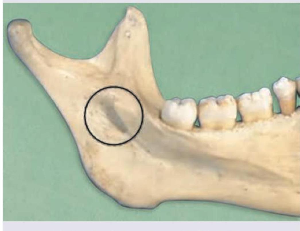
Which nerve passes through the structure shown? (Recent NEET Pattern 2019)

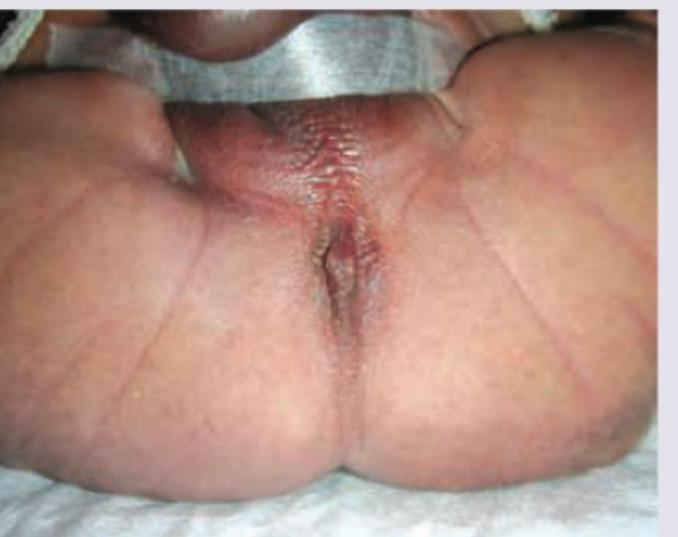
All investigations are useful in work up of this condition except:

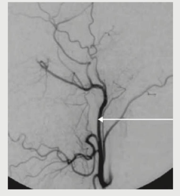
What is the name of the marked blood vessel, which is a branch of external carotid artery?

Testicular artery is a branch of -
Left anterior descending artery is a direct branch of
Highest point of iliac crest is seen at?
Which of the following is TRUE about bronchopulmonary segments?
Most prominent and largest air cell of ethmoidal sinus?
Articular surfaces ≥3 are present in which type of joint?
Explanation: ***T10*** - The dermatome for **T10** corresponds to the level of the **umbilicus**. - This is a crucial anatomical landmark for dermatomal assessment, neurological examination, and clinical correlation. - The T10 dermatome is consistently tested in NEET-PG examinations. *T12* - The dermatome for **T12** is located just inferior to the umbilicus, typically corresponding to the **suprapubic region** or inguinal area. - It does not correspond to the umbilicus itself. *L1* - The dermatome for **L1** is generally located in the **inguinal region** and upper anterior thigh. - This is significantly lower than the umbilicus. *L3-L4* - **L3-L4** refers to a **vertebral level**, not a dermatomal level. - While the umbilicus is anatomically located at approximately the L3-L4 vertebral level, the question asks for the **dermatome**, which is T10. - Dermatomes and vertebral levels should not be confused.
Explanation: ***Inferior alveolar nerve*** - The image points to the **mandibular foramen**, an opening on the medial surface of the mandibular ramus. - The **inferior alveolar nerve** enters the mandible through this foramen to supply sensation to the mandibular teeth. *Lingual nerve* - The **lingual nerve** typically runs anterior to the inferior alveolar nerve in the infratemporal fossa but does not pass through the mandibular foramen. - It supplies general sensation and taste to the anterior two-thirds of the tongue. *Buccal nerve* - The **buccal nerve** passes between the two heads of the lateral pterygoid muscle and supplies sensation to the buccal mucosa and gingiva, not traveling within the mandible. - It arises from the anterior division of the mandibular nerve. *Hypoglossal nerve* - The **hypoglossal nerve (CN XII)** is a motor nerve for the tongue muscles and is located entirely outside the mandible. - It exits the skull through the hypoglossal canal and runs in the neck and floor of the mouth.
Explanation: ***Urine for meconium particles*** - **Urine for meconium particles** is actually a valuable and standard investigation in **anorectal malformations** to detect **rectourinary fistulas**. - The presence of meconium in urine indicates a **communication between the rectum and urinary tract**, which is crucial information for surgical planning and management. *Invertogram at 24 hours* - An **invertogram** is a plain X-ray taken with the infant inverted for 3-5 minutes, allowing gas in the rectum to rise to the highest point. - This helps determine the **level of rectal pouch termination** relative to the **pubococcygeal line**, which is essential for distinguishing between high and low anorectal malformations. *Ultrasound* - **Perineal ultrasound** can assess the **perineal body thickness**, presence of a **rectal pouch**, and help visualize **fistulous connections**. - It is particularly useful in **real-time assessment** of the anatomy and can identify associated **genitourinary anomalies** commonly seen with anorectal malformations. *Lateral pelvic radiography* - A **lateral pelvic radiograph** provides information about the **sacral anatomy** and helps assess the **level of anorectal malformation**. - It is crucial for identifying associated **sacral anomalies** (sacral dysgenesis, hemisacrum) which occur in up to 60% of patients with anorectal malformations and affect surgical outcomes.
Explanation: ***Correct: Lingual artery*** - The image shows a vessel branching off the anterior aspect of the **external carotid artery** and extending towards the tongue region, which is characteristic of the **lingual artery**. - This artery typically arises at the level of the **greater horn of the hyoid bone** and supplies the **tongue** and floor of the mouth. - The anterior projection and course towards the tongue region are key identifying features. *Incorrect: Superior thyroid artery* - The superior thyroid artery typically branches off the **external carotid artery** more inferiorly, often near its origin, and descends to supply the **thyroid gland**. - This vessel, in contrast, is marked higher up and projects anteriorly towards the tongue, not inferiorly towards the thyroid. *Incorrect: Ascending pharyngeal artery* - The **ascending pharyngeal artery** usually arises from the medial or posterior aspect of the external carotid artery as a small, slender branch. - It ascends vertically to supply the **pharynx**, prevertebral muscles, and middle ear, not showing the anterior horizontal course seen in the marked vessel. *Incorrect: Maxillary artery* - The maxillary artery is a terminal branch of the **external carotid artery** that originates behind the neck of the mandible, deep to the parotid gland at a higher level. - It has a complex course with many branches supplying deep structures of the face, but its origin is much more posterior and superior than the marked vessel.
Explanation: ***Abdominal aorta*** - The **testicular arteries**, also known as **gonadal arteries**, originate directly from the anterior aspect of the **abdominal aorta**. - They typically arise just inferior to the **renal arteries** at the level of the second lumbar vertebra (L2) and descend to supply the testes. *Common iliac artery* - The common iliac artery is a terminal branch of the **abdominal aorta**, but it gives rise to the internal and external iliac arteries, not directly the testicular artery. [2] - It bifurcates at the level of the sacroiliac joint. [3] *External iliac artery* - The external iliac artery primarily supplies the **lower limb** and gives off the inferior epigastric and deep circumflex iliac arteries. [1] - It does not directly provide branches to the testes. *Internal iliac artery* - The internal iliac artery primarily supplies the **pelvic organs**, gluteal region, and perineum. - While it has numerous branches, none of them are the main gonadal arteries; it contributes to the blood supply of the reproductive organs through other smaller branches. [3]
Explanation: ***Left coronary artery*** - The left coronary artery (LCA) is a major coronary artery that arises from the **aorta** and quickly branches into two main arteries: the **left anterior descending (LAD) artery** [1] and the circumflex artery. - The LAD artery, also known as the **"widowmaker"**, supplies oxygenated blood to the **anterior wall of the left ventricle** and the interventricular septum, making it crucial for heart function. *Right coronary artery* - The **right coronary artery (RCA)** typically supplies the **right atrium**, most of the **right ventricle**, and the inferior wall of the left ventricle, which are distinct areas from the LAD's supply. - The RCA originates from the **right sinus of Valsalva** and travels in the atrioventricular groove, while the LAD originates from the left main coronary artery [1]. *Circumflex artery* - The circumflex artery is another main branch of the **left coronary artery**, typically supplying the **lateral and posterior walls of the left ventricle** and the left atrium. - While it branches from the same parent vessel as the LAD, it is a direct branch itself, not the origin of the LAD [1]. *Ascending aorta* - The ascending aorta is the initial part of the aorta that originates from the **left ventricle** and gives rise to the **coronary arteries** (both left and right coronary arteries). - It is the source from which the **left coronary artery** (and thus the LAD indirectly) originates, but it is not a direct branch itself.
Explanation: ***L4*** - The **highest point of the iliac crest** typically corresponds to the level of the **L4 vertebral body**. - This anatomical landmark is crucial for procedures like **lumbar punctures** and determining the location for **epidural anesthesia**. *L3* - The L3 vertebral level is generally located slightly **above the highest point of the iliac crest**. - While close, it is not the most consistent anatomical correlation for the highest point. *S2* - The **S2 vertebral level** is significantly **below the iliac crests**, marking the approximate midpoint of the sacroiliac joint. - This level is used as a landmark for the **dermatome of the posterior thigh**. *S1* - The **S1 vertebral level** is also located **below the iliac crests**, forming the most superior segment of the sacrum. - It is used as a landmark for the **dermatome of the lateral foot and posterior leg**, and corresponds to the ankle jerk reflex.
Explanation: Detailed knowledge of lung anatomy is essential for thoracic surgery. ***Pulmonary veins are intersegmental*** - The **pulmonary veins** run in the connective tissue septa **between** the bronchopulmonary segments, making them **intersegmental** [1]. - This anatomical arrangement allows surgeons to ligate the veins without affecting adjacent segments during a segmentectomy. *Spherical in shape* - Bronchopulmonary segments are typically **pyramid-shaped** or cone-shaped, with their apices directed towards the hilum [1]. - Their irregular, wedge-like structure allows them to fit together within the lung. *Artery is intersegmental* - The **bronchial arteries** and **pulmonary arteries** are **intrasegmental**, meaning they run **within** the bronchopulmonary segment alongside the bronchi. - Each segment has its own arterial supply, allowing for independent blood flow. *Non resectable* - Bronchopulmonary segments are considered **functionally and surgically independent units**, making them **resectable** [1]. - This allows for the removal of diseased segments while preserving healthy lung tissue, such as in cases of **lung cancer** or localized infection.
Explanation: ***Bulla ethmoidalis*** - The **bulla ethmoidalis** is universally present and is consistently described as the **largest and most prominent** of the anterior ethmoid air cells. - It forms an anatomical landmark that is consistently superior and posterior to the **hiatus semilunaris**. *Onodi cell* - The **Onodi cell** is a posterior ethmoid air cell that pneumatizes laterally and superiorly into the sphenoid bone, in close proximity to the optic nerve. - While clinically significant due to its relationship with the optic nerve, it is not the largest or most prominent ethmoidal air cell. *Haller cell* - A **Haller cell** is an infraorbital ethmoid cell located along the floor of the orbit, extending into the maxillary sinus. - These cells can contribute to **ostial obstruction** of the maxillary sinus but are typically small compared to the bulla ethmoidalis. *Agger nasi cell* - The **agger nasi cell** is the most anterior ethmoid air cell, located in the lacrimal bone anterior to the frontal recess. - It is often one of the first ethmoid cells to pneumatize but is generally small and not considered the most prominent.
Explanation: ***Compound joint*** - A **compound joint** is defined by having **three or more articular surfaces**, meaning multiple bones articulate within a single joint capsule. - Examples include the **elbow joint** (humerus, ulna, radius) and the **knee joint** (femur, tibia, patella). *Complex joint* - A **complex joint** is characterized by the presence of an **intra-articular disc or meniscus**, which divides the joint cavity. - This feature helps to absorb shock and improve congruity, but it doesn't necessarily dictate the number of articular surfaces. *Synarthrosis joint* - A **synarthrosis joint** is an **immovable joint** where bones are very close together, such as the sutures of the skull. - These joints prioritize stability over movement and typically do not involve distinct articular surfaces in the same way as synovial joints. *Simple joint* - A **simple joint** involves the articulation of **only two bones** or two articular surfaces within a single joint capsule. - Examples include the **phalangeal joints** (interphalangeal joints) and the **hip joint** (femur and pelvis).
Skeletal System
Practice Questions
Articular System
Practice Questions
Muscular System
Practice Questions
Cardiovascular System
Practice Questions
Lymphatic System
Practice Questions
Nervous System
Practice Questions
Respiratory System
Practice Questions
Digestive System
Practice Questions
Urinary System
Practice Questions
Reproductive System
Practice Questions
Endocrine System
Practice Questions
Integumentary System
Practice Questions
Get full access to all questions, explanations, and performance tracking.
Start For Free